Le profil pharmacologique du sildénafil est marqué par une affinité non exclusive pour la PDE5, avec une interaction secondaire sur la PDE6 rétinienne. Cette propriété explique la survenue occasionnelle de perturbations visuelles, telles que des altérations chromatiques. Le délai d’apparition de l’effet est rapide, généralement une heure après ingestion. Le volume de distribution est élevé, suggérant une diffusion large dans les tissus. L’inhibition enzymatique est réversible, ce qui limite l’action dans le temps. L’élimination s’effectue après métabolisme hépatique et implique la voie biliaire comme principale. Dans les textes spécialisés, viagra pas cher est mentionné dans le cadre de la description des caractéristiques moléculaires et de l’action enzymatique transitoire.
Microsoft powerpoint - poster biowin aderenne_format coupé.pptx
The FTIR spectrum of prostate cancer cells signs the mode of action The FTIR spectrum of prostate cancer cells si of anticancer drugs gns the mode of action
Allison Derenne, Régis Gasper and Erik Goormaghtigh
Laboratory of Structure and Function of Biological Membranes, Center for Structural Biology and Bioinformatics, Université Libre de Bruxelles (ULB)
E-mail: [email protected] This work was supported by the FNRS
OBJECTIVES 3) PLS PLS D iscr Discriminant model OBJECTIVES
A discriminant PLS model was built from the 50 pixels means using the spectral range 1764 – 1300 cm-1.
Traditionally, new drugs are evaluated for their potential to kill cancer cell lines. This approach is obviously not
This method determines a linear combination of variables that separates as much as possible the different
sufficient, and molecules with new modes of action are required. We suggest here that the infrared spectrum of cells
classes of samples. Discrimination rules are established in order to minimize within groups separation
exposed to anticancer drugs could offer an opportunity to obtain a fingerprint of the metabolic changes induced by
and maximize separation between groups.
these drugs. Because the infrared spectrum of cells yields a precise image of all the chemical bonds present in thesample, different drug actions are likely to each yield a unique fingerprint characteristic of the “mode of action” of the
Predicted
therapeutical agent under investigation. Topoisomerases Antimetabolites Antimicrotubules AIM OF THE PROJECT : USE INFRARED SPECTROSCOPY AS A NEW SCREENING METHOD inhibitors SELECTING NEW ANTICANCER AGENTS ACCORDING TO THEIR MODE OF ACTION Topoisomerases MATERIALS AND METHODS inhibitors Antimetabolites Antimicrotubules Table 1: Percentage (%) of correctly classified mean spectra (means of 50 pixels) by the PLS model built on the1764-1300 cm-1 spectral range. About 3000 spectra not involved in the building of the model were used in this testset.
Application of this PLS model to individual pixels reveals some causes of the misclassifications
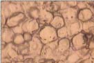
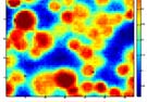
Antimicrotubules Antimetabolites Topoisomerases inhibitors Filtered out Fig 1 : Cells were cultured in the absence or presence of the 7different drugs for 48 hrs (1). Cells were detached with 0.05%trypsin/EDTA, washed 3 times with an isotonic solution (NaCl0.9%) (2), re-suspended at ca 50,000 cells/µl and laid downAntimicrotubules 2 window (3) then an IR image (4096Antimetabolites pixels) was recorded between 3900 and 800 cm-1 (4).Topoisomerases inhibitors
and are located in the correct class. Filtered out 1) Determination of IC50
The concentration of drug reducing cell growth by 50% after 72 hrs (IC ) was determined by a MTT test for
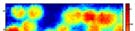
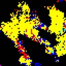
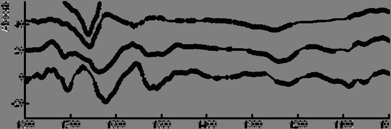
Fig 3 : Application of the PLS to a PC-3 cell culture exposed to doxorubicin (A) and mercaptopurine (B). The color of describes the result of the classification. Black pixels have been eliminated by the filters (see Methods).POTENTIAL APPLICATIONS AND KEY BENEFITS 2) Drug-induced spectral modification of the PC-3 prostate cell line New selected molecule inhibitor inhibitor Fig 4: Hierarchical classification of the difference spectra presented in figure 3.Fig 2: Differences between mean spectra for PC-3 cells exposed to a drug as indicated in the right margin and the
This method allows to rapidly classify the mode of action of potential drugs and could improve the efficiency
control. A Student t-test was computed at every wavenumber with a significance level of α=0.005. Each markedCONCLUSIONS : IN THIS WORK, WE OBTAINED SPECIFIC FTIR SPECTRAL SIGNATURES FOR THE EFFECT OF 7 ANTICANCER MOLECULES ON PROSTATE TUMOR PC-3 CELLS. FURTHERMORE, THE SIGNATURE OF DRUGS WITH SIMILAR MODES OF ACTION ARE VERY CLOSE. THIS STUDY SUGGESTS THAT FTIR IMAGING PROVIDES AN UNIQUE SIGNATURE OF CELLULAR PATHWAYS TRIGGERED BY THE MOLECULE TESTED AND IN TURN COULD OFFER AN OBJECTIVE SELECTION CRITERION FOR NEW DRUGS WITH ORIGINAL MODES OF ACTION.
___________________________________________________________________________ CLINICAL GUIDELINES for SUBCUTANEOUS INFUSION (HYPODERMOCLYSIS) Clinical Policy Folder Ref No: 16 APPROVED BY: Policy and Guideline Ratification Group (PGRG) Date of Issue: July 2010 Version No: 1.3 Date of review: May 2012 Author: Alison Griffiths. Matron District Nursing NHS South Glouces
A) Drug-Induced Allergies 1) KRAFT, D. und L. WIDE: Clinical patterns and results of radioallergosorbent-test (RAST) and 2) KRAFT, D., D. WILSON and M. E. DEVEY: Penicillin allergy studies by a modified red cell linked Int. Archs. Allergy Appl. Immunol. 52, 248-256 (1976). 3) KRAFT, D., A. ROTH, P. MISCHER, H. PICHLER and H. EBNER: Specific and total serum IgE measurements in the diagnosis


 The FTIR spectrum of prostate cancer cells signs the mode of action