Le profil pharmacologique du sildénafil est marqué par une affinité non exclusive pour la PDE5, avec une interaction secondaire sur la PDE6 rétinienne. Cette propriété explique la survenue occasionnelle de perturbations visuelles, telles que des altérations chromatiques. Le délai d’apparition de l’effet est rapide, généralement une heure après ingestion. Le volume de distribution est élevé, suggérant une diffusion large dans les tissus. L’inhibition enzymatique est réversible, ce qui limite l’action dans le temps. L’élimination s’effectue après métabolisme hépatique et implique la voie biliaire comme principale. Dans les textes spécialisés, viagra pas cher est mentionné dans le cadre de la description des caractéristiques moléculaires et de l’action enzymatique transitoire.
Femininebeauty.info
Original Contribution O R I G I NA L C O N T R I BU T I O N The combination of 2% 4-hydroxyanisole (mequinol) and 0.01% tretinoin effectively improves the appearance of solar lentigines in ethnic groups Zoe Diana Draelos, MD
While the efficacy and safety of topical 4-hydroxyanisole (mequinol) 2%/tretinoin 0.01%therapy has been established in Caucasian populations, those with skin types I–II, littleresearch has focused on individuals with darker skin types. The purpose of this open-labelstudy was to evaluate the efficacy and safety of mequinol 2%/tretinoin 0.01% solutionin the treatment of solar lentigines in Asian, Latin/Hispanic, and African Americanethnic groups with skin types II–V. Subjects were required to have ≥ 10 solar lentigineson the dorsal forearms/hands and ≥ 3 on the face. One lesion was designated the targetlesion, however, all lesions were treated. Patients were treated with topical mequinol 2%/tretinoin 0.01% and clinically evaluated at 4, 8, 12, 16, 20, and 24 weeks as well as4 weeks following treatment cessation. At each visit, lesions were evaluated using Targetand Overall Lesion Pigmentation Index scores ranging from 0 (lightest) to 8 (darkest),where 4 indicated equal pigment with surrounding skin. Efficacy was determined basedon pigmentation index scores, and safety was assessed using laboratory monitoringand adverse event (AE) reporting. Over 80% of the 259 subjects completing this studyresponded to mequinol 2%/tretinoin 0.01% therapy, with a majority of subjects main-taining clinical benefit at 4 weeks post-treatment. Most AEs reported were tolerable andoverall mequinol 2%/tretinoin 0.01% therapy had a favorable benefit-to-risk ratio. Thisstudy therefore supports the theory that topical mequinol 2%/tretinoin 0.01% is aneffective and safe treatment of solar lentigines in ethnic populations, and in those withdark skin types. Keywords:ethnic groups, mequinol, solar lentigines, tretinoin
exposure. Lesions occur most frequently in fair-skinned
Introduction
individuals and in middle-aged and elderly populations.1–3
Solar lentigines are a common dermatologic condition
This condition currently causes significant cosmetic
that manifest as localized, hyperpigmented, macular
concerns for more than 20 million Americans and is
lesions usually found on sun-exposed areas of the skin.
becoming increasingly prevalent due to the increased rate
The benign condition is caused by an increased number
at which the US population is aging.4 Although new and
of active melanocytes and increased melanin production
effective therapies are available, the majority of therapeutic
in response to chronic, accumulated ultraviolet radiation
research has focused on Caucasian populations or thosewith skin types I or II (Table 1). This is not surprising given the
Correspondence: Zoe Diana Draelos, MD, 2444 North Main Street, High
epidemiological data on the disease. However, the condition
Point, NC 27262. E-mail: [email protected]
affects all skin types and requires research supporting its
Accepted for publication May 16, 2006
treatment in ethnic groups and those with darker skin.5–7
2006 Blackwell Publishing • Journal of Cosmetic Dermatology, 5, 239– 244
Mequinol 2%/tretinoin 0.01% use with ethnic groups • Z D DraelosTable 1 Demographic information.
With the exception of these investigations, no studies have
specifically addressed solar lentigines in non-Caucasian
Given the limited exploration in these patient popula-
tions, the present study was designed to assess the efficacy
and safety of mequinol 2%/tretinoin 0.01% therapy in
II – Usually burns, tans less than average
The present study was an open-label, single-arm safety
III – Sometimes burns (mildly), tans about average
and efficacy study of mequinol 2%/tretinoin 0.01%
IV – Rarely burns, tans more than average
topical solution in the depigmentation of circumscribed
macular solar lentigines in males and females ≥ 30 years
of Asian, Latin/Hispanic, African American ethnicity andskin types II–V. Using the wand applicator to specifically
target only the solar lentigines, subjects applied thequick-drying test solution twice daily to lesions on the
Recent evidence supports the use of combination therapy
face, forearms, and hands for up to 24 weeks.
with mequinol 2%/tretinoin 0.01% (Solagé®, Barrier
After giving written informed consent, 45 (17.4%)
Therapeutics, Princeton, NJ) as an effective and safe treat-
males and 214 (82.6%) females (total n = 259) were
ment for solar lentigines.1–4,8,9 Widely accepted treatments,
enrolled into 17 study centers in the United States and 3
such as tretinoin and hydroquinone monotherapies, and
in Canada. The mean age of the subjects was 55.8 years,
ablative treatment strategies, such as cryo- and laser
with a range of 31–82 years. Sixty-three (24.3%) of the
removal, often produce variable depigmentation results.
subjects were Asian, 35 (13.5%) were African American,
Additionally, such therapies often have negative side
and 161 (62.2%) were Hispanic (Table 1). Eligible subjects
effects and can result in significant patient discomfort.3
were demonstrably nongravid and had ≥ 10 solar lentigines
Mequinol 2%/tretinoin 0.01% combines two effective
on the dorsal forearms/hands and ≥ 3 on the face. A single
depigmenting agents and has been proven more effective
lesion in each area was designated as a target lesion,
than either agent alone while offering a safety profile
however, all lesions were treated. At baseline the overall
similar to tretinoin monotherapy.2 Tretinoin (all-trans-
lesion pigmentation index score was 6 on a 9-point scale
retinoic) is a vitamin A analogue thought to inhibit
ranging from 0 (lightest) to 8 (darkest), where 4 (equal)
melanogenesis through growth factor modulation,3,10 The
indicated equal pigmentation with the surrounding skin
exact mechanism of action of mequinol’s depigmentation
effects is unknown although it is speculated that it
Subjects returned to the clinic for evaluations after 4,
includes oxidation by tyrosinase to cytotoxic products in
8, 12, 16, 20 and 24 weeks of treatment and again
melanocytes, a direct/selective toxic effect on melanocytes,
4 weeks later for follow-up assessment. In this open-label,
or inhibition of melanin formation.11–15
single-arm study, all the assessments for a subject were
Although multiple recent studies have shown mequinol
performed by the same investigator; evaluations were
2%/tretinoin 0.01% combination therapy to provideclinically significant improvement in up to 80% of
Table 2 Lesion pigmentation index.
patients, little work has been done to specifically evaluate
its efficacy in ethnic populations or in those with
0 – Extremely lighter than pigment of surrounding skin
darker skin tones.2,3 One study has suggested that 2%
hydroquinone-cyclodextrin therapy significantly reduced
1 – Markedly lighter than pigment of surrounding skin
pigmentation in Asian patients with solar lentigines,
2 – Moderately lighter than pigment of surrounding skin
another study explored concomitant application of all-trans-
3 – Slightly lighter than pigment of surrounding skin
retinoic acid aqueous gel with hydroquinone-lactic acid
4 – Equal with pigment of surrounding skin
5 – Slightly darker than pigment of surrounding skin
ointment for bleaching of senile lentigines and post-
6 – Moderately darker than pigment of surrounding skin
inflammatory hyperpigmentations, while other work has
7 – Markedly darker than pigment of surrounding skin
looked at hyperpigmentation responses to topical whiten-
8 – Extremely darker than pigment of surrounding skin
ing agents in women of South-east Asian descent.5–7
2006 Blackwell Publishing • Journal of Cosmetic Dermatology, 5, 239– 244
Mequinol 2%/tretinoin 0.01% use with ethnic groups • Z D Draelos
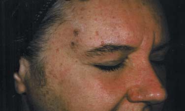
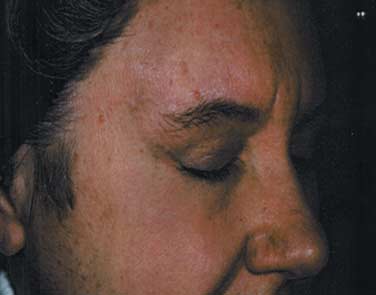
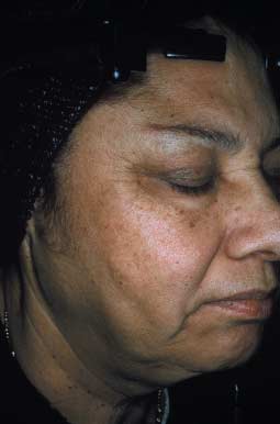
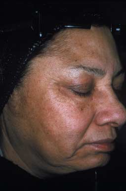
based on the target lesions presented at baseline. Basedon investigator interest, experience and training, colorphotographs, for illustrative purposes only, were takenat three selected study sites at baseline, week 24, orupon successful depigmentation to grade 4 (Fig. 1a,b). Figure 2a,b is representative before and after images ofsubjects with deeply pigmented skin who were treatedwith the test formulation. At each visit, a Target and anOverall Lesion Pigmentation Index Score were recordedbased on clinical assessment for each of the two treatmentareas, resulting in a total of four scores per visit. Preg-nancy status was monitored at each visit, and laboratorytests (blood chemistry, urinalysis, and hematology) wereperformed at baseline, 12 weeks, and 24 weeks (or at lastvisit) for safety monitoring. For each treated lesion, if ascore of 4 was reached, treatment was stopped for thatlesion. When all treated lesions were at grade 4, treatmentwas stopped altogether, regardless of study duration.
Efficacy was determined by clinical assessment using
Target Lesion and Overall Lesion Pigmentation Index scores and was quantified by dividing responders into three categories: • Complete responders – subjects who reached an Overall • Partial responders – subjects who had significant
improvement in their pigmentation, defined as animprovement of at least 1 grade, compared to baseline;
• Treatment failures – subjects who did not fulfill the Figure 1 Facial pigmentation lightening occurred in the facial
above criteria or subjects achieving an Overall Lesion
treatment sites after 24 weeks of therapy in an African American
Pigmentation Index score of less than 4.
subject. (a) baseline and (b) 24 weeks. Figure 2 Representative (a) before and (b) after images of subjects with deeply pigmented skin who were treated with the test formulation.
2006 Blackwell Publishing • Journal of Cosmetic Dermatology, 5, 239– 244
Mequinol 2%/tretinoin 0.01% use with ethnic groups • Z D Draelos
The time to reach a response in partial and complete
Table 3 Summary of adverse events.
responders was defined as the time from first treatmentapplication to the first measurable response.
In addition to the aforementioned laboratory tests,
safety was assessed by evaluating reported adverse events(AEs). Assessment of AEs, including abnormal pigmentation
changes, was performed at each visit, and any events were
recorded throughout the duration of the study. Subjects
were instructed to avoid sun exposure as well as exposure
to other sources of ultraviolet radiation (i.e., tanning beds)
whenever possible for the duration of the study. They
were also instructed to keep treatment areas covered with
Over 80% of subjects responded to treatment as assessed
by Overall Lesion Pigmentation Index scores. One
hundred and seventy-six (68.0%) and 198 (76.4%)
subjects had partial responses for the arm and face,
respectively, while 35 (13.5%) and 21 (8.1%) subjects
experienced complete responses for the same respective
areas. Forty-eight (18.5%) and 40 (15.4%) subjects had
treatment failures for the arm and face, respectively.
Over 85% of subjects responded to treatment based on
Target Lesion Pigmentation Index scores. Once again the
A subject was counted only once per AE regardless of the number of
majority of subjects had a partial response to treatment
with 176 (68.0%) and 185 (71.4%) showing a lesion
*Defined as erythema plus at least two additional, prespecified
improvement by at least 1 grade for the arm and face
signs/symptoms (scaling, dryness, stinging/burning) starting at
regions, respectively. Forty-nine (18.9%) and 45 (17.4%)
subjects experienced complete responses, while 34 (13.1%)and 29 (11.2%) subjects had treatment failures for thearm and face, respectively.
AEs were reported by 121 (46.7%) subjects, all of whom
The median time to response (partial and complete) for
had dermatological AEs. Thirteen (5%) subjects discon-
the Overall Lesion Pigmentation Index was 56 days
tinued treatment due to an AE, nine (3.5%) of which were
(range 21–173 days) for both the arm and face. Median
dermatological in nature. Additionally, 15 (5.8%) subjects
response times were 51 and 35 days for the Target Lesion
stopped treatment due to dermatological AEs in one area
Pigmentation Index for the same respective regions. At
(either the face or arm) but remained in the study and
4-week follow-up, the majority of subjects maintained
continued undergoing treatment in the nonaffected area.
the same pigmentation index scores as observed at the
Serious AEs were reported by six (2.3%) subjects; all were
nondermatological, including one subject who died of a
Exposure to the study medication ranged from 1 to
myocardial infarction considered unrelated to study
232 days (mean of 151.2 days) with most subjects
treatment by the investigators. Laboratory AEs were
(> 70% per area) receiving treatment for over 141 days.
infrequent; hematuria occurred in nine (3.5%) subjects
Mequinol 2%/tretinoin 0.01% treatment showed an
(seven of which were female). No laboratory AE resulted
overall favorable safety profile consistent with previous
in treatment discontinuation and none were considered
clinical studies.1–3 Of 259 subjects, 160 (61.8%) reported
related to treatment by the investigators.
1 or more AEs; 125 (48.3%) subjects reported 1 or moredermatological AEs. Thirty-two (12.4%) subjects reported
Conclusions
hypopigmentation or halo-hypopigmentation (12 [4.6%]and 20 [7.7%], respectively) (Table 3). The majority of
Overall, mequinol 2%/tretinoin 0.01% combination
these events (84%) resolved during the study. Drug-related
therapy demonstrated a favorable risk-to-benefit ratio in
2006 Blackwell Publishing • Journal of Cosmetic Dermatology, 5, 239– 244
Mequinol 2%/tretinoin 0.01% use with ethnic groups • Z D Draelos
the treatment of solar lentigines in Asian, Latin/Hispanic,
and irritation response—as they may have affected efficacy
and African American subjects with skin types II–V.
This study has demonstrated that over 80% of treated
The barrier properties of skin can produce variations in
subjects achieved a significant response to the therapy
skin permeability and thus absorption. When skin types
for both arm and facial lesions, the majority of which
V and VI was compared to types II and III, researchers
maintained clinical benefit 4 weeks post-treatment. These
concluded that darker skin was more compact than
results mirror efficacy findings previously reported for
lighter skin probably due to more cornified cell layers,
light-skinned individuals and provide new evidence
and that darker skin showed greater epidermal barrier.21
supporting the use of mequinol 2%/tretinoin 0.01%
These results support the findings of an earlier study that
specifically in ethnic and dark-skinned populations.2 It
found that lighter skin was more permeable to certain
is important to note that mequinol 2%/tretinoin 0.01%
was found to have favorable treatment response times
In the 1980s and early 1990s, researchers studied
(both partial and complete) comparable, if not better,
racial and ethnic differences in irritant reaction to topically
than those seen with other therapies.16,17 The median
applied chemicals by using biologic parameters. Based on
response time for the Overall Lesion Pigmentation Index
findings from several studies, Berardesca et al.23–27 con-
was 56 days (range 21–173 days) for both the arm and
face; median response times were 51 and 35 days for the
• black subjects’ skin displays a stronger skin irritant
Target Lesion Pigmentation Index for the same respective
regions. Noting the realistic amount of time it may take
• the skin of black subjects is more sensitive to irritants
for a noticeable difference when treating solar lentigines
may help in the management of patient expectations for
• black subjects display less erythema, less blood vessel
therapy and ultimately support compliance.
reactivity, and less cutaneous blood flow to irritants
There are no racial differences in the number of
melanocytes,18,19 but the actual number of melanocytes
• Hispanic subjects show a stronger irritant reaction
differ from one person to another or from one area of the
compared with white subjects; their irritant reaction is
body to another,20 which may account for the differences
in treatment response of the test drug. All treatment
• Hispanic subjects have stronger irritant reactions when
modalities have nonresponders. The small proportion of
subjects in this trial with no improvement is not surprising
• Hispanic and white subjects have similar erythematous
and was probably due to the individual’s unique biologic
makeup. The approximately 3% difference in response
Many current treatment strategies for solar lentigines
with treated facial and arm lesions is consistent with the
are associated with either inadequate depigmentation
results found in studies of light-skinned subjects and
responses or unfavorable side effects resulting in severe
most likely not linked to racial differences2,3 but rather to
patient discomfort.3 It has been widely reported that
the difference in size and distribution of melanosomes in
formulations containing hydroquinone and/or glucocor-
ticoids are associated with an increased risk of ochronosis,
Although AEs were reported by a significant number of
a paradoxical hyperpigmentation response.28 Such
patients, the treatment was overall tolerable, especially
adverse reactions can be alarming to patients who initially
when considering the overwhelming treatment response
sought treatment in order to resolve hyperpigmentation
rate. The most commonly reported AEs were erythema,
abnormalities. Through avoidance of undesirable
skin discomfort, and halo-hypopigmentation, occurring
side effects such as ochronosis, mequinol 2%/tretinoin
in 24.7%, 20.1%, and 7.7% of subjects, respectively.
0.01% offers patients an attractive alternative to other
These results are significantly more favorable than AE
therapies being used to treat solar lentigo. Therefore, the
results previously reported in light skin populations
present study supports the use of mequinol 2%/tretinoin
further supporting the use of mequinol 2%/tretinoin
0.01% combination therapy as an effective, tolerable
treatment option for ethnic and dark-skinned subjects.
Because of the inherent ethnic differences in skin biology,
it was important to target a specific population of subjectswith darker skin. Although the efficacy and safety results
References
of this trial were similar to those of earlier studies1–3 that
1 Farris PK. Combination therapy for solar lentigines. J Drugs
comprised a majority of light-skinned subjects, two differ-
Dermatol 2004; 3: S23–S26.
ences in ethnic skin biology were of concern—absorption
2 Fleisher AB, Schwartzel EH, Colby SI et al. The
2006 Blackwell Publishing • Journal of Cosmetic Dermatology, 5, 239– 244
Mequinol 2%/tretinoin 0.01% use with ethnic groups • Z D Draelos
combination of 2% 4-hydroxyanisole (mequinol) and
lipid peroxidation in the tyrosinase/4-hydroxyanisole
0.01% tretinoin is effective in improving the appearance
system: possible mechanism of killing of malignant
of solar lentigines and related hyperpigmented lesions in
melanoma cells by 4-hydroxyanisole. Biochem Int
two double-blind multicenter clinical studies. J Am Acad
1992; 26: 397– 403. Dermatol 2000; 42: 459 – 67.
15 Katsambas A, Antoniou C. Melasma. Classification and
3 Ortonne JP, Camacho F, Wainwright N, Bergfelt L,
treatment. J Eur Acad Dermatol Venereol 1995; 4: 217–23.
Westerhof W, Roseeuw D. Safety and efficacy of combined
16 Food and Drug Administration. (2005) Tri-Luma Cream.
use of 4-hyroxyanisole (mequinol) 2%/tretinoin 0.01%
solution and sunscreen in solar lentigines. Cutis 2004; 74:
label/2002/21112lbl.pdf (Accessed 2 September 2005).
17 Grimes PE. A microsponge formulation of hydroquinone 4%
4 1996 US Government Census Report, Washington, DC:
and retinol 0.15% in the treatment of melasma and
postinflammatory hyperpigmentation. Cutis 2004; 74:
5 Hermanns JF, Petit L, Piérard-Franchimont C, Paquet P,
Piérard GE. Assessment of topical hypopigmenting agents
18 Szabo G. Pigment cell biology. In: M Gordon, ed.
on solar lentigines of Asian women. Dermatology 2002;
Mitochondria and Other Cytoplasmic Inclusions. New York:
204: 281–6.
6 Petit L, Piérard GE. Analytic quantification of solar
19 Staricco RS, Pinkus H. Quantitative and qualitative data on
lentigines lightening by a 2% hydroquinone-cyclodextrin
the pigment cells of adult human epidermis. J Invest
formulation. J Eur Acad Dermatol Venereol 2003; 17: Dermatol 1957; 28: 33– 45.
20 Toda K, Patnak MA, Parrish A, Fitzpatrick TB, Quevedo WC.
7 Yoshimura K, Harii K, Aoyama T, Iga T. Experience with a
Alteration of racial differences in melanosome distribution
strong bleaching treatment for skin hyperpigmentation in
in human epidermis after exposure to ultraviolet light. Nat
Orientals. Plast Reconstr Surg 2000; 105: 1097–110. New Biol 1972; 236: 143–4.
8 Colby SI, Schwartzel EH, Huber FJ et al. A promising new
21 Reed JT, Ghadially R, Elias PM. Skin type, but neither race
treatment for solar lentigines. J Drugs Dermatol 2003; 2:
nor gender, influence epidermal permeability barrier
function. Arch Dermatol 1995; 131: 1134 – 8.
9 Jarrat M. Mequinol 2%/tretinoin 0.01% solution: An
22 Wedig JH, Maibach HI. Percutaneous penetration of
effective and safe alternative to hydroquinone 3% in the
dipyrithione in men: effect of skin color (race). J Am Acad
treatment of solar lentigines. Cutis 2004; 74: 319–22. Dermatol 1981; 5: 433– 8.
10 Akdeniz N, Calka O, Ozbek H, Metin A. Anti-inflammatory
23 Berardesca E, Maibach HI. Racial differences in sodium
effects of tretinoin (all-trans-retinoic acid) 0.1% and
lauryl sulfate induced cutaneous irritation: black and
adapalene 0.1% in rats. Clin Exp Dermatol 2005; 30:
white. Contact Dermatitis 1988; 18: 65–70.
24 Berardesca E, Maibach HI. Sodium-lauryl-sulphate-
11 Bessou S, Pain C, Taieb A. Use of human skin reconstructs
induced cutaneous irritation comparison of white and
in the study of pigment modifiers. Arch Dermatol 1997; 133:
Hispanic subjects. Contact Dermatitis 1988; 19: 136– 40.
25 Berardesca E. Racial differences in skin function. Acta Derm
12 Belcher HJ, Nizam M, O’Neill TJ. Intra-arterial
Venereol 1994; 185 (Suppl.): 44 – 6.
4-hydroxyanisole chemotherapy for locally recurrent
26 Wilson D, Berardesca E, Maibach HI. In vitro
malignant melanoma: a re-appraisal. Br J Plastic Surg
transepidermal water loss: differences between black and
1992; 45: 208 –10.
white human skin. Br J Dermatol 1988; 119: 647–52.
13 Garcia Reverte JM, Bernabeu Esclapez A, Munoz Ramos J,
27 Berardesca E, Maibach HI. Sensitive and ethnic skin: a need
Vicente Ortega V, Canteras Jordana M. Effects of various
for special skin care agent? Dermatol Clin 1991; 9: 89– 92.
antineoplastic agents on an established human melanoma
28 Bongiorno MR, Arico M. Exogenous ochronosis and striae
cell line (G-361). Histol Histopathol 1995; 10: 79– 84.
atrophicae following the use of bleaching creams. Int J
14 Koga S, Nakano M, Ito T, Tomita Y. Importance of iron in
Dermatol 2005; 44: 112 –5.
2006 Blackwell Publishing • Journal of Cosmetic Dermatology, 5, 239– 244
DR. ZIV CENTER FOR ORTHOPAEDIC SPECIALISTS Hand / Wrist / Elbow Postoperative Program PLEASE READ! Keep the hand elevated above the level of your heart as much as possible, especially for the first 48-72 hours. A sling is good while standing/ going out for 1 week. After a week the sling is not needed and it is best to use it as little as possible. If you are sitting on a couch, take the s
Copyright � The Korean Academy Inflammatory Myofibroblastic Tumor on Intercostal Nerve Presentingas Paraneoplastic Pemphigus with Fatal Pulmonary InvolvementInflammatory myofibroblastic tumors (IMTs) are benign neoplasms that can occurat different anatomic sites with nonspecific clinical symptoms. A 48-yr-old womanpresented with a 2-month history of a relapsed oral ulcer, progressive dysp
 Mequinol 2%/tretinoin 0.01% use with ethnic groups • Z D Draelos
based on the target lesions presented at baseline. Basedon investigator interest, experience and training, colorphotographs, for illustrative purposes only, were takenat three selected study sites at baseline, week 24, orupon successful depigmentation to grade 4 (Fig. 1a,b).
Mequinol 2%/tretinoin 0.01% use with ethnic groups • Z D Draelos
based on the target lesions presented at baseline. Basedon investigator interest, experience and training, colorphotographs, for illustrative purposes only, were takenat three selected study sites at baseline, week 24, orupon successful depigmentation to grade 4 (Fig. 1a,b).