Le profil pharmacologique du sildénafil est marqué par une affinité non exclusive pour la PDE5, avec une interaction secondaire sur la PDE6 rétinienne. Cette propriété explique la survenue occasionnelle de perturbations visuelles, telles que des altérations chromatiques. Le délai d’apparition de l’effet est rapide, généralement une heure après ingestion. Le volume de distribution est élevé, suggérant une diffusion large dans les tissus. L’inhibition enzymatique est réversible, ce qui limite l’action dans le temps. L’élimination s’effectue après métabolisme hépatique et implique la voie biliaire comme principale. Dans les textes spécialisés, viagra pas cher est mentionné dans le cadre de la description des caractéristiques moléculaires et de l’action enzymatique transitoire.
Appnote2.indd
A P P L I C A T I O N N O T E #2 Laser Capture Microdissection of muscle fi ber populations and expression analysis by RT-PCR Sven Fraterman and Neal Rubinstein, Department of Cell and Developmental Biology, University of Pennsylvania School of Medicine, Philadelphia Abstract
acetylcholine esterase stain by Karnowsky and
To locate gene products in distinct muscle
with a mouse-monoclonal slow myosin heavy
allotypes, a novel protocol was established.
SIFs show a large, C-shaped neuromuscular
junction after acetylcholine esterase staining
and do not react with the anti-slow MyHC
immunohistochemistry was used to provide
criteria for their selective isolation by laser
of MIFs is smaller and more circular and
capture microdissection while preserving
immuno-staining. Since mRNA is sensitive
reaction showed a differential expression
pattern of muscle specifi c genes in different
the slides to dehydration by xylenes was
muscle fi bers in laser captured material. Introduction
traditionally been the paradigm for studies
Molecular Probes ZenonTM technology.
atypical muscle groups such as extraocular
This protocol describes a way to isolate
groups (called “allotypes”) have been the
subject of research for the past decades. The limb and EOM allotypes differ in
Equipment and Reagents Equipment and Labware
fi bers and 80% of EOM fi bers are singly
innervated fi bers (SIFs) which have only
one neuromuscular junction. About 20% of
EOM fi bers are multiply-innervated fi bers
(MIFs): they have multiple neuromuscular
junctions2. In the past, immunohistochemistry
used to study differences between muscle
fiber populations3,4, While useful, these
techniques are limited in their capability
mixed muscle fi ber population. Moreover,
To overcome these problems, laser capture
microdissection was used to isolate different
multiple gene products by polymerase chain
reaction. The muscle fi ber populations in
6 5 0 . 9 6 2 . 3 0 2 0 t e l 8 8 8 . 4 6 6 . 7 9 1 1 t o l l - f r e e 6 5 0 . 9 6 2 . 3 0 3 9 f a x t e c h s u p p o r t @ a r c t u r . c o m w w w . a r c t u r . c o m
A P P L I C A T I O N N O T E #2
required for RNA isolation and analysis by RT-PCR:
Incubation oven Microcentrifuge Arcturus alignment tray Thermo Cycler with heated lid Horizontal gel chamber Power supply Gel Doc 2000TM Documentation
RNase-free Technique
In addition to the usual precautions listed below, some special precautions were taken connected to the rapid staining protocol: 1. Use RNase® AWAY according to
the manufacturer’s instructions on laboratory bench surfaces, cryostat, cryostat knife, PixCell® II Laser Capture Microdissection System and Arcturus alignment tray.
2. Disposable gloves are to be frequently
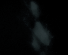
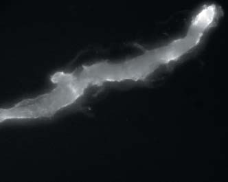
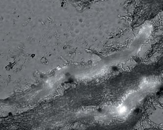
Figure 1. Laser Capture Microdissection of Muscle Fiber Populations. A multiply innervated muscle fi ber is shown before (A) and after (B) laser capture mircodissection. The fi ber shows
strong immunohistochemical staining for slow myosin and the two grappe-like neuromuscular junctions are
Figures C and D show the laser capture microdissection process of a population of singly innervated muscle fi bers. This
preparation of all staining solutions and
fi ber population is negative for slow MyHC immuno-staining and show comparable large neuromuscular junctions
which are again marked with an arrow. Figures A and C are taken with the fl uorescence and dimmed visual light on
and Figures B and D show the fi ber after microdissection on the laser microdissection cap with only fl uorescence
light on. Some autofl uoresence is detectable in Figure D, but the signal is weaker than in Figure B.
preserving the capability to distinguish
different muscle fi ber populations.
• 5 mg of acetylthiocholine iodide are
2. The muscle is cut into 10 µm sections
the staining solution has to be prepared
fresh since the acetylthiocholine starts
process, the slides with 4 sections each
are stored on dry ice and are afterwards
3. For the acetylcholine esterase stain by
6 5 0 . 9 6 2 . 3 0 2 0 t e l 8 8 8 . 4 6 6 . 7 9 1 1 t o l l - f r e e 6 5 0 . 9 6 2 . 3 0 3 9 f a x t e c h s u p p o r t @ a r c t u r . c o m w w w . a r c t u r . c o m
A P P L I C A T I O N N O T E #2
with the prepared staining solution for 20 minutes.
8. After 20 minutes, the slide is washed
twice for 3 minutes in DEPC treated water. Figure 2. Semi-quantitative RT-PCR of different muscle fi ber populations.
Gel pictures of polymerase chain reaction for epsilon- and gamma- subunit of the acetylcholine receptor, slow
minute in a fl ow hood the slide is ready
Muscle fi bers were distinguished by their reactivity to the acetylcholine esterase stain by Karnowsky and Roots and
to Alexa Fluor 488 conjugated slow MyHC antibody. In the leg, three different populations were analyzed: (NMJ),
the neuromuscular junction region of fi bers negative for slow MyHC; (slow +), non innervated region of fi bers
positive for slow MyHC; (slow -), non innervated region of fi bers negative for slow MyHC. In the EOMs, fi bers were
again distinguished by their reactivity to anti-slow MyHC antibody and their staining for acetylcholine esterase.
(NMJ-SIF), the neuromuscular junction region of fi bers negative for slow MyHC - these are all singly innervated;
(NMJ-MIF), the neuromuscular junction region of fi bers positive for slow MyHC - these are all multiply-innervated;
(slow +), non innervated region of fi bers positive for slow MyHC - i.e. MIFs; (slow -), non innervated region of fi bers
11. The microdissection laser is set to 7
the desired tissue was captured based on
the fl uorescence signal (Figures 1B and
12. SIFs were dissected based on their large
17. 15 µl of each PCR product were loaded
described in its protocol within 2 hours
The data presented in this application note
demonstrates that it is possible to isolate a
single muscle fi ber type or a distinct muscle
fi ber population and analyze from it the
expression of muscle specifi c genes by RT-
esterase stain. To dissect single fi bers, it
PCR. Figure 1 demonstrates the isolation of an
can be advantageous to search for fi bers
individual fi ber type by LCM. The ZenonTM
staining of slow MyHC positive fi bers gives
picking up undesired fi ber populations.
a strong, unambiguous signal in less than
16. To study expression of muscle specifi c
35 minutes. The acetylcholine esterase stain
13. To avoid cross-contamination by other
of Karnowsky and Roots allows us to assess
cell types, every cap is scanned visually
rapidly the innervation pattern of muscle
fi bers. When used alone, this esterase stain
on a fresh slide and viewing it at lowest
can provide results within 5-10 minutes.
power. This is followed by a scan at the
Hence, rapid identifi cation of distinct fi bers
6 5 0 . 9 6 2 . 3 0 2 0 t e l 8 8 8 . 4 6 6 . 7 9 1 1 t o l l - f r e e 6 5 0 . 9 6 2 . 3 0 3 9 f a x t e c h s u p p o r t @ a r c t u r . c o m w w w . a r c t u r . c o m
A P P L I C A T I O N N O T E #2
Table I. PCR Cycling Conditions, Reaction Components and Volumes Used
and innervation pattern allows us time to process tissue via LCM and isolate mRNA
FastStart PCR Master Mix Thermal Cycler programming
without signifi cant time for degradation.
Using this type of analysis on limb muscle
confi rms data previously obtained by other
means4. Hence, we can apply the technique
to another muscle allotype, the EOM, and
be sure that this protocol and the Arcturus
PixCell II Laser Capture Microdissection
System is suffi cient to distinguish muscle fiber types and give new insight into
Table II. PCR Primer Sequences and Annealing Temperatures
Forward primer (fwd) Annealing References size (bp) Reverse primer (rev) Physiol. Genomics. 2002;9:71-84.
Immunohistochemical identifi cation of slow-tonic fi bers in human extrinsic eye muscles. Invest. Ophthalmol. Vis. Sci. 1979;18:303-6.
distribution of myosin heavy chain isoforms among rat extraocular muscle fi ber types. Invest. Ophthalmol. Vis. Sci. 2000;41:3391-8.
B. Imprinting of acetylcholine receptor messenger RNA accumulation in mammalian neuromuscular synapses. Nature. 1990;344:544-7.
5. Karnowsky MJ, Roots, L.A. A “direct
coloring” thiocholine method for cholinesterase. J. Histochem. Cythochem. 1964:219-221.
S, Nadal-Ginard B, Rubinstein NA, Kelly AM. Slow myosin in developing rat skeletal muscle. J. Cell. Biol. 1987;104:447-59.
6 5 0 . 9 6 2 . 3 0 2 0 t e l 8 8 8 . 4 6 6 . 7 9 1 1 t o l l - f r e e 6 5 0 . 9 6 2 . 3 0 3 9 f a x t e c h s u p p o r t @ a r c t u r . c o m w w w . a r
Leser-Rezensionen Ein Buch, das Hoffnung schenkt Ich bin Realist und konnte mir gar nicht vorstellen das Saft und Tee diesen Erfolg haben könnten. Aber selbst an Krebs erkrankt greift man nach jedem Strohhalm. Heute kann ich Ihnen sagen, dass ich diese Kur immer wieder als Regenerations-Kur mache. Der damit verbundene Salbeitee tut mir gut. Ich trinke ihn schon zwei Jahre und bei mir und mei
[· · · ] a transformational programming method-ology that includes a fully operational set-the-oretic proof checker [· · · ][· · · ] examples of two moderately difficult pro-gram derivations. One of these derives a highlevel form of an algorithm to compute the bisim-ulation equivalence relation [· · · ] the other de-rives an algorithm to minimize the number ofstates in a deterministi
 A P P L I C A T I O N N O T E #2
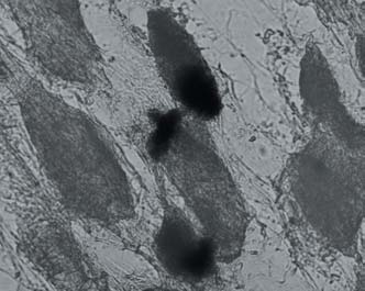
 A P P L I C A T I O N N O T E #2
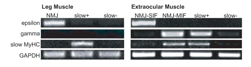 A P P L I C A T I O N N O T E #2
 A P P L I C A T I O N N O T E #2