030515 identification of severe acute respiratory syndrome in canada
The new england journal of medicine
Identification of Severe Acute Respiratory
Susan M. Poutanen, M.D., M.P.H., Donald E. Low, M.D., Bonnie Henry, M.D.,
Sandy Finkelstein, M.D., David Rose, M.D., Karen Green, R.N.,
Raymond Tellier, M.D., Ryan Draker, B.Sc., Dena Adachi, M.Sc.,
Melissa Ayers, B.Sc., Adrienne K. Chan, M.D.,
Danuta M. Skowronski, M.D., M.H.Sc., Irving Salit, M.D., Andrew E. Simor, M.D.,
Arthur S. Slutsky, M.D., Patrick W. Doyle, M.D., M.H.Sc., Mel Krajden, M.D.,
Martin Petric, Ph.D., Robert C. Brunham, M.D., and Allison J. McGeer, M.D.,
for the National Microbiology Laboratory, Canada,
and the Canadian Severe Acute Respiratory Syndrome Study Team*
From the Toronto Medical Laboratoriesand Mount Sinai Hospital Department of
b a c k g r o u n d
Microbiology, Toronto (S.M.P., D.E.L., K.G.,A.J.M.); the Department of Laboratory Med-
Severe acute respiratory syndrome (SARS) is a condition of unknown cause that has re- icine and Pathobiology (S.M.P., D.E.L., R.T.,
cently been recognized in patients in Asia, North America, and Europe. This report sum- A.E.S., A.J.M.), Department of Medicine Di-marizes the initial epidemiologic findings, clinical description, and diagnostic findings vision of Infectious Diseases (D.E.L., A.K.C.,
that followed the identification of SARS in Canada.
Medicine and Interdepartmental Divisionof Critical Care (A.S.S.), University of To-
ronto, Toronto; the City of Toronto Public
SARS was first identified in Canada in early March 2003. We collected epidemiologic, Health Department (B.H.); Scarborough
Hospital, Toronto (S.F., D.R.); the Hospital
clinical, and diagnostic data from each of the first 10 cases prospectively as they were for Sick Children, Toronto (R.T., R.D., D.A.,
identified. Specimens from all cases were sent to local, provincial, national, and inter- M.A.); Epidemiology Services (D.M.S.) andnational laboratories for studies to identify an etiologic agent.
Laboratory Services (M.K., M.P.), British Co-lumbia Centre for Disease Control, Van-
couver; University Health Network, Toronto(I.S.); Sunnybrook and Women’s College
The patients ranged from 24 to 78 years old; 60 percent were men. Transmission oc- Health Sciences Centre, Toronto (A.E.S.);
curred only after close contact. The most common presenting symptoms were fever (in St. Michael’s Hospital, Toronto (A.S.S.); the100 percent of cases) and malaise (in 70 percent), followed by nonproductive cough (in Department of Pathology and Laboratory
100 percent) and dyspnea (in 80 percent) associated with infiltrates on chest radiogra- Sciences Centre and University of British
phy (in 100 percent). Lymphopenia (in 89 percent of those for whom data were avail- Columbia, Vancouver (P.W.D.); and the Uni-able), elevated lactate dehydrogenase levels (in 80 percent), elevated aspartate amino- versity of British Columbia Centre for Dis-
ease Control, Vancouver (R.C.B.) — all in
transferase levels (in 78 percent), and elevated creatinine kinase levels (in 56 percent) Canada. Address reprint requests to Dr.
were common. Empirical therapy most commonly included antibiotics, oseltamivir, McGeer at the Toronto Medical Laborato-and intravenous ribavirin. Mechanical ventilation was required in five patients. Three ries and Mount Sinai Hospital, Department
of Microbiology, 600 University Ave., Rm.
patients died, and five have had clinical improvement. The results of laboratory inves- 1460, Toronto, ON M5G 1X5, Canada.
tigations were negative or not clinically significant except for the amplification of hu-man metapneumovirus from respiratory specimens from five of nine patients and the *Members of the National Microbiology
isolation and amplification of a novel coronavirus from five of nine patients. In four
Team groups are listed in the Appendix. c o n c l u s i o n s
This article was published at www.nejm.orgon March 31, 2003.
SARS is a condition associated with substantial morbidity and mortality. It appears tobe of viral origin, with patterns suggesting droplet or contact transmission. The role of N Engl J Med 2003;348:1995-2005. human metapneumovirus, a novel coronavirus, or both requires further investigation. Copyright 2003 Massachusetts Medical Society.
Downloaded from nejm.org on November 19, 2012. For personal use only. No other uses without permission.
Copyright 2003 Massachusetts Medical Society. All rights reserved.
The new england journal of medicine
She died three days later, on March 5, at home, nine
(SARS) is a condition of unknown cause that days after the onset of her illness. An autopsy was
shas recently been recognized in patients in not performed.
Asia, North America, and Europe. As defined by the
The index patient’s 43-year-old son (Patient 2),
World Health Organization (WHO), a suspected who had an underlying history of type 2 diabetescase is disease in a person with a documented fever and hypertension, had fever and diaphoresis on(temperature, >38°C), lower respiratory tract symp- February 27, two days after his mother first notedtoms, and contact with a person believed to have symptoms. Within approximately five days he be-had SARS or a history of travel to a geographic area came afebrile, but concurrently, a nonproductivewhere there has been documented transmission cough, chest pain, and dyspnea developed. A chestof the illness. A suspected case that involves chest radiograph revealed moderate air-space disease inradiographic findings of pneumonia, acute res- the right middle and lower lobes, for which he re-piratory distress syndrome (ARDS), or an unex- ceived antibiotics. Because of persistent symptoms,plained respiratory illness resulting in death with he was assessed at a hospital and noted to have a fe-autopsy results demonstrating the pathology of ver (temperature, 39.8°C) and an oxygen saturationARDS without an identifiable cause is considered of 82 percent while breathing room air. A chest ra-a probable case.1
diograph revealed bibasilar air-space disease. He
This report summarizes the initial epidemio- was admitted to the hospital with a diagnosis of
logic findings, clinical description, and diagnostic community-acquired pneumonia and was support-findings that followed the identification of SARS in ed with noninvasive ventilation and treated withCanada.
broad-spectrum antibiotics. Antituberculous medi-cation and airborne precautions were added whentuberculosis was considered after the first day of his
admission. Contact precautions were also added,
d e s c r i p t i o n o f t h e o u t b r e a k
given uncertainty about the underlying infectious
agent. By day 2 of his admission, his respiratory sta-
The first cases in Toronto were linked to members tus had deteriorated, and he was intubated and re-of a multigenerational family of Hong Kong de- ceived mechanical ventilation. Despite intensivescent who live in Toronto (Fig. 1 and 2). The To- physiological support, multiorgan dysfunction syn-ronto index case (Patient 1) and her husband trav- drome developed, and he died on March 13, 2003,eled to Hong Kong to visit relatives from February 6 days after admission, and 15 days after becoming13 through February 23, 2003. While in Hong ill. All routine investigations for etiologic agentsKong visiting their son, Patient 1 and her husband were negative. At autopsy, the lung tissue revealedstayed at Hotel A from February 18 through Feb- diffuse alveolar damage consistent with pathologicruary 21. Another hotel guest, who eventually was manifestations of ARDS. Intraalveolar and inter-identified as the source patient for SARS in Hong stitial mononuclear cells suggesting a possible viralKong, also stayed on the same floor at Hotel A.2 cause were also noted, but no viral cytopathic effectPatient 1 and her husband stayed in the hotel only was seen. Examination of the liver revealed micro-at night, spending the days visiting their son. They vesicular fatty change, focal hemorrhages, andreturned to their apartment in Toronto, which hepatocyte necrosis with scattered acidophilicthey shared with two sons, a daughter-in-law, and a bodies, but no viral inclusions were seen. Thefive-month-old grandson (Household A), on Feb- spleen showed large areas of probable ischemicruary 23, 2003.
necrosis and some atypical lymphocytes in periar-
Patient 1, a 78-year-old woman with a history of teriolar sheaths. Further information on the evalu-
type 2 diabetes and coronary heart disease, had fe- ation for specific pathogens is given below. ver, anorexia, myalgias, a sore throat, and mild non-
On March 8 and 9, because of concern about
productive cough two days after returning home. possible tuberculosis in the family, the remainingThree days later, her family physician noted pharyn- five adult family members and their three childrengeal erythema but no other abnormalities on physi- (5 months old, 9 years old, and 17 years old), whocal examination. An oral antibiotic was prescribed, had all been exposed to the index patient, underwentand she was sent home. Two days later, she noted screening chest radiography. All had fever, cough,the development of increasing cough with dyspnea. dyspnea, or all three, as well as abnormal chest ra-
Downloaded from nejm.org on November 19, 2012. For personal use only. No other uses without permission.
Copyright 2003 Massachusetts Medical Society. All rights reserved.
s e v e r e a c u t e r e s p i r a t o r y s y n d r o m e i n c a n a d a
Figure 1. Pedigree of the Epidemiologically Linked Toronto Patients with SARS.
Shading indicates suspected or probable cases of SARS, and slashes patients who died.
diographs, except for the three children and the 2 and his wife (Patient 4) on March 6, when theyhusband of Patient 3, who were and continue to be were both symptomatic. Patient 7 had a severe head-asymptomatic with normal chest radiographs. SARS ache on March 9, followed by fevers (temperatureswas considered to be a possible explanation for of up to 40°C), myalgias, and malaise. Four days lat-these abnormalities and for the deaths of Patient 1 er, a nonproductive cough developed, and she wasand Patient 2, who in retrospect met the criteria for noted to have fever (temperature, 38.5°C) and tach-probable SARS. (Patient 1 met almost all of the cri- ypnea with an oxygen saturation of 100 percent onteria, although an autopsy and microbiologic inves- room air. Chest radiography revealed a subtle lefttigations were not completed to rule out identifiable basilar infiltrate. She was admitted to a medicalcauses.) In the light of this possible explanation, ward with a diagnosis of suspected SARS and haseach of the symptomatic adults was reassessed on subsequently recovered, coincident with receivingMarch 13. One met the criteria for suspected SARS broad-spectrum antibiotics, oseltamivir, and intra-(Patient 4), and three met the criteria for probable venous ribavirin. SARS (Patient 3, Patient 5, and Patient 6). All four
The second additional identified case was in a
were admitted to the hospital, three of them to in- 76-year-old man of non-Asian descent (Patient 8)tensive care units; one patient required mechanical who had a history of type 2 diabetes, coronary heartventilation. All four were treated with broad-spec- disease, and hypertension and who was evaluatedtrum antibiotics, oseltamivir, and intravenous riba- at the hospital to which Patient 2 was admitted. Pa-virin and have recovered fully, with the exception of tient 8 was assessed in the emergency departmenttwo who continue to have mild dyspnea on exertion on March 7 for atrial fibrillation and observed over-approximately three weeks after the onset of their night on a gurney separated by a cotton curtain 1 toillness.
2 m from Patient 2, who was being held overnight
As a result of media attention, three additional without respiratory or contact precautions awaiting
cases of SARS were identified. The first case was in an inpatient hospital bed. Patient 8 was dischargeda previously healthy 37-year-old female family phy- home on March 8, and two days later he had feversician of Asian descent (Patient 7) who saw Patient (temperatures of up to 40°C), diaphoresis, and fa-
Downloaded from nejm.org on November 19, 2012. For personal use only. No other uses without permission.
Copyright 2003 Massachusetts Medical Society. All rights reserved.
The new england journal of medicine
Incubation V Requirement for mechanical ventilation Patient 1 (78-year-old woman) Patient 2 (43-year-old man) Patient 3 (38-year-old woman) Patient 4 (24-year-old woman) Patient 5 (34-year-old man) Patient 6 (79-year-old man) Patient 7 (37-year-old woman)
Exposed to Patient 2 and Patient 4 in an outpatient clinic
Patient 8 (76-year-old man)
Exposed to Patient 2 in an emergency room
Patient 10 V V V V V V V V V V V V V V (55-year-old man) Figure 2. Timeline of Events in the Epidemiologically Linked Canadian Cases of SARS.
tigue. A chest radiograph revealed right-upper-lobe his return to Toronto on March 14, he was admittedand bibasilar interstitial infiltrates; despite antibi- to the hospital with a diagnosis of probable SARSotic treatment, a nonproductive cough and worsen- and was treated with broad-spectrum antibiotics,ing dyspnea subsequently developed, along with oseltamivir, and intravenous ribavirin. Two days af-hypothermia (temperature, 36.6°C) and an oxygen ter admission, his respiratory status worsened andsaturation of 70 percent on room air. He was ad- he required intubation and ventilation. His condi-mitted to an intensive care unit with a diagnosis of tion has since stabilized and is slowly improving,probable SARS and required intubation and venti- but he continues to require ventilatory support 16lation. Despite receiving broad-spectrum antibiot- days after the onset of his illness. ics, oseltamivir, intravenous ribavirin, and intensivesupport, he died on March 21, 5 days after admis- Vancouversion and 12 days after the onset of his illness. An au- The only case in Vancouver was in Patient 10, a 55-topsy was performed, the results of which were year-old, previously healthy man who traveled withpending at this writing.
his wife to Hong Kong and Bali from February 20
The third additional case (in Patient 9) was unre- through March 6, 2003. While visiting Hong Kong
lated to this family cluster. He is a 62-year-old man from February 20 to February 24, Patient 10 and hisof non-Asian descent with a history of atrial fibrilla- wife also stayed in Hotel A, but on a different floortion who had chest pain, sore throat, and light- from Patient 1, Patient 6, and the hotel guest whoheadedness, followed by cough, dyspnea, and fever, was eventually identified as the source patient forwhile traveling in Southeast Asia in early March. On SARS in Hong Kong. When in the hotel, Patient 10
Downloaded from nejm.org on November 19, 2012. For personal use only. No other uses without permission.
Copyright 2003 Massachusetts Medical Society. All rights reserved.
s e v e r e a c u t e r e s p i r a t o r y s y n d r o m e i n c a n a d a
and his wife did not eat at a common dining facility,nor did they entertain or visit other guests in the
Table 1. Clinical Features of the Canadian Patients with SARS at Presentation.
hotel. While traveling, two days after leaving HongKong, Patient 10 had malaise followed two days
Variable
later by fever (temperature, 39.4°C), chills, and head-ache. Progressive dyspnea and a nonproductive
cough developed a day later. After returning to Van-
couver on March 7, he was assessed and found to
have a temperature of 38.5°C, an oxygen saturation
Symptoms
of 45 percent on room air, and mixed air-space and
reticular opacification diffusely on chest radiogra-phy. He was admitted to the intensive care unit, and
within 24 hours he required intubation and ventila-
tion to maintain adequate oxygenation. He was
soon recognized as having probable SARS and hasbeen treated with broad-spectrum antibiotics. He
remains in intensive care on ventilatory support 30
days after the onset of his illness. s u m m a r y o f c l i n i c a l f e a t u r e s a n d i n i t i a l i n v e s t i g a t i o n s
A summary of the clinical features and initial inves-
tigations of the first 10 cases of SARS identified in
Investigations
Canada (8 probable and 2 suspected) is given in Ta-ble 1. All of the patients were adults, ranging from
24 to 78 years of age. Six of the 10 were men. Eight
of the 10 were of Asian descent. Three had a diag-
nosis of type 2 diabetes mellitus (Patient 1, Patient
2, and Patient 8); two had underlying pulmonary
Lymphopenia (cell count <1.5¬10 /liter)
disease (asthma in Patient 3 and chronic cough of
unclear cause in Patient 6); and four had a history
of smoking (Patient 2, Patient 6, Patient 8, and Pa-
tient 9) although none still smoked. Given the pa-
tients who had a defined exposure time (Patient 3,
Patient 7, and Patient 8), the incubation period can
be estimated to range from 3 to 10 days. However,
Alanine aminotransferase (>1.5¬ upper
we are unable to exclude the possibility of a one-
The presenting symptoms included fever in all
cases and nonspecific symptoms such as malaise(7 of 10 cases) and myalgias (2 of 10 cases). Three * Although all 10 patients had a history of fever at presen-
tation to the hospital, on examination only 5 of 9 were fe-
of the 10 patients had chest pain, 3 had sore throat,
brile (temperature, 38.4 to 40°C); 1 had a low-grade fever
and 3 had headache as part of their initial presenta-
(temperature, 37.9°C), and 3 had hypothermia (tempera-
tion. Although a nonproductive cough (in all 10
cases) and dyspnea (8 of 10 cases) were common,these respiratory symptoms were not the present-ing symptoms in 5 cases. In three patients the fevers
On presentation to the hospital, five of nine
had improved by the time respiratory symptoms patients were febrile (temperature, 38.4 to 40°C),occurred. Five of the 10 patients had diarrhea, and one had a low-grade fever (temperature, 37.9°C),1 had vomiting, although 4 of these patients were and three had hypothermia (temperature, 35.5 toalso taking medications frequently associated with 36.5°C). Tachycardia (in five of nine cases), tachyp-gastrointestinal side effects. No patient had a rash. nea (in seven of nine), and borderline low blood
Downloaded from nejm.org on November 19, 2012. For personal use only. No other uses without permission.
Copyright 2003 Massachusetts Medical Society. All rights reserved.
The new england journal of medicine
pressure (in five of nine) were common. Oxygensaturation while breathing room air was less than
95 percent in seven of nine patients. Physical exam-ination was normal in all patients outside the respi-ratory system. Crackles were noted at the basessymmetrically (in three of nine patients) or asym-metrically (in two of nine). Bronchial breath soundsor egophony were noted in three patients. Chest ra-diographs revealed abnormalities in all of the ninepatients who underwent radiography. In all butthree patients, changes were bilateral and predomi-nantly in the basal lung zones. Abnormalities weresubtle at first in five of the nine patients, primarilyinvolving a reticular interstitial pattern. For two of
these patients, subsequent chest radiographs wereread as normal. All other patients had progressivesymmetric involvement of predominantly air-spacedisease on subsequent radiographs. Pleural effu-sions were not seen. A representative series of chestradiographs is shown in Figure 3.
noted included lymphopenia, elevated lactate dehy-drogenase levels, elevated aspartate aminotransfer-ase levels, and elevated creatine kinase levels. Otherless common abnormalities included mild throm-
c a s e m a n a g e m e n t
All seven patients who presented to the hospital inToronto and were recognized as having suspectedor probable SARS were treated empirically with rec-ommended doses of oral oseltamivir and broad-spectrum antibiotics, as well as intravenous ribavi-rin in the dosing schedule recommended for thetreatment of viral hemorrhagic fever (a loading doseof 2 g, followed by 1 g every six hours for four days,then 500 mg every eight hours for another four tosix days).3 The three other patients were treated with
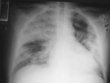
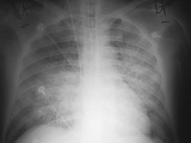
Figure 3. The Course of Disease in Patient 8.
empirical broad-spectrum antibiotics alone. All cas-
Patient 8, a 76-year-old man who was exposed to Patient 2
es were managed with use of respiratory and contact
on March 7, had fever (temperatures of up to 40°C), dia-phoresis, and fatigue three days later on March 10. A
precautions as soon as the diagnosis of suspected
chest radiograph obtained on March 14 revealed right-
or probable SARS had been considered.
upper-lobe and bibasilar interstitial infiltrates (Panel A). Despite antibiotic treatment, he subsequently noted a nonproductive cough and increasing dyspnea and was
c l i n i c a l c o u r s e
Five of the patients with SARS required mechanical
admitted to an intensive care unit on March 16. A chest radiograph obtained on March 17 revealed bilateral
ventilation for worsening respiratory failure at one
patchy air-space disease with relative sparing of the right
point in their illness, and two of these patients have
lung base and left upper lobe (Panel B), and the patient
died (Patient 2 and Patient 8). Patient 2 initially re-
was intubated and received mechanical ventilation for
ceived noninvasive ventilation and was then intu-
respiratory distress. Progressive respiratory failure and
bated and received mechanical ventilation until his
worsening of chest-radiography findings occurred on March 20 (Panel C), and the patient died on March 21.
death four days later. Tidal volumes were 500 to
Downloaded from nejm.org on November 19, 2012. For personal use only. No other uses without permission.
Copyright 2003 Massachusetts Medical Society. All rights reserved.
s e v e r e a c u t e r e s p i r a t o r y s y n d r o m e i n c a n a d a
700 ml, and minute ventilation largely ranged be- ognized as having SARS and in whom an autopsy tween 10 and 15 liters per minute. Peak inspiratory was not performed.) pressures ranged from 17 to 42 cm of water but were generally less than 35 cm of water, with posi- histopathological investigations tive end-expiratory pressure levels of 5 to 15 cm of Autopsy tissue from Patient 2 was subjected to im- water. The ratio of the partial pressure of oxygen munohistochemical tests for influenzaviruses A (PaO ) to the fraction of inspired oxygen (FiO ) in and B, respiratory syncytial virus, adenovirus, Hen-
the first 96 hours was generally more than 150, but dra and Nipah viruses, hantavirus, measles virus,it decreased thereafter to between 100 and 150; the enterovirus, flaviviruses, Old World arenavirus, ty-PaO never dropped below 71 mm Hg. Patient 8 was phus and spotted fever rickettsia, coxiella species,
intubated and received ventilation with tidal vol- Yersinia pestis, Mycoplasma pneumoniae, and Chlamydo-umes of 500 to 750 ml. The FiO ranged from 0.9 to phila (Chlamydia) pneumoniae. All were negative.
1.0, with PaO :FiO ratios of 56 to 85 despite posi-
tive end-expiratory pressure levels of 14 cm of water. microbiologic investigations Peak inspiratory pressures generally ranged from Bacterial and Fungal Examination 24 to 38 cm of water, and the partial pressure of car- Routine bacterial and fungal examination was com- bon dioxide increased from about 25 to about 50 pleted on all blood, respiratory, and urine specimens mm Hg over a period of four days despite increases from 9 of the 10 patients, yielding negative results. in minute ventilation from about 15 to 20 liters per Specifically, cultures as well as direct examination minute. Although both patients had markedly ab- (where appropriate) were completed on all blood, normal PaO :FiO ratios, they were never extreme- respiratory, and urine specimens received, yielding
ly hypoxemic and probably did not die of severe hy- negative results. In addition, cultures for legionellapoxemia.
species, direct fluorescent antibody testing against
Patient 1 also died at home, increasing the total legionella species on all respiratory specimens re-
number of deaths to 3 among the 10 patients. All of ceived, and testing for the presence of Legionella pneu-the deaths occurred in patients who had an underly- mophila serogroup 1 antigen in urine specimens re-ing immunocompromised state (type 2 diabetes).
Of the three patients who were treated with
To date, bacterial molecular testing has been
broad-spectrum antibiotics alone, two died and one completed on all respiratory specimens receivedremains in intensive care requiring mechanical ven- from 6 of the 10 patients, yielding negative results. tilation. Of the seven patients who were treated with Specifically, DNA was extracted, and polymerase-intravenous ribavirin and oral oseltamivir in addi- chain-reaction (PCR) detection for targets specifiction to broad-spectrum antibacterial therapy, one for L. pneumophila, M. pneumoniae, C. pneumoniae,died and one remains in intensive care requiring C. psittaci, Chlamydophila at the genus level, Y. pestis,mechanical ventilation but with signs of clinical Bacillus anthracis, and 16S rRNA was negative. improvement. The other five, including one whohad required mechanical ventilation, showed im- Virologic Examinationprovement within the first five days of treatment. Routine direct virologic examination of all respira-They have all since recovered fully, with the excep- tory and stool specimens received from 9 of the 10tion of two who remain mildly dypsneic on exer- patients was completed, yielding negative results. tion approximately three weeks after the onset of This included negative electron-microscopical ex-their illness.
amination and negative direct fluorescent antibodytesting against influenzaviruses A and B, parainflu-
l a b o r a t o r y i n v e s t i g a t i o n s
enzaviruses 1, 2, and 3, adenovirus, and respiratory
Histopathological and microbiologic investigations syncytial virus in all specimens, with the exceptionof specimens received were conducted at local, na- of one subsequently unconfirmed positive directtional, and international laboratories. Histopatho- fluorescent antibody result for influenzavirus B in alogical testing was completed on autopsy tissue specimen from Patient 10. from Patient 2. Routine and specialized microbio-
Viral molecular testing has been completed on
logic investigations were completed on all speci- all respiratory and blood specimens received frommens received in 9 of the 10 cases. (No specimens 9 of the 10 patients. Specifically, DNA was extractedwere sent from Patient 1, who died before being rec- and PCR was completed for targets specific to vari-
Downloaded from nejm.org on November 19, 2012. For personal use only. No other uses without permission.
Copyright 2003 Massachusetts Medical Society. All rights reserved.
The new england journal of medicine
ous DNA viruses, yielding negative results for ade- fied from all five cultures. In addition, nestednoviruses, parvoviruses, circoviruses, herpesviruses, RT-PCR using the same primers plus 5'TGTTA-and orthopoxviruses. In addition, RNA was extract- AACCAGGTGGAAC3' and 5'CCTGTGTTGTA-ed and reverse-transcription–PCR (RT-PCR) was GATTGCG3' amplified the coronavirus directlycompleted for targets specific to various RNA virus- from bronchoalveolar-lavage fluid from three ofes, including influenzaviruses A and B, respiratory nine patients tested, all of whom also had coronavi-syncytial virus, parainfluenzavirus subtypes 1, 2, 3, rus isolated from cell culture as described above. and 4, human metapneumovirus, filoviruses (Ebola
At a different laboratory, a coronavirus was also
and Marburg viruses), arenaviruses, measles virus, identified independently by amplification directlymumps virus, Hanta viruses, and Crimean–Congo from bronchoalveolar-lavage fluid from three of sixhemorrhagic fever virus, yielding negative results.
patients tested. All three of these patients had coro-
Further virologic studies were completed on all navirus isolated from cell culture and amplification
respiratory specimens received from 9 of the 10 pa- as described above. Reverse transcription was com-tients. These included viral cultures (including in- pleted using the primer 5'GCATAGGCAGTAGTT-oculation onto cell culture and into embryonated GCATC3', followed by PCR targeting a highly con-hen eggs and intracerebral inoculation of suckling served region of the coronavirus polymerase genemice), immune electron microscopy of nasopharyn- with use of the primer pair 5'TGATGGGATG-geal swabs and bronchoalveolar fluids with serum GGACTATCCTAAGTGTGA3' and 5'TTGCATCAC-obtained during the convalescent phase from Pa- CACTAGTTGTGCCACCAGGTT3'. One of thetient 10, RT-PCR for conserved portions of the poly- amplicons was sequenced (GenBank accessionmerase gene of RNA viruses, and nested RT-PCR number AY271716), and although the nucleotide se-with genus-specific degenerative primers for para- quence was different from that of any known coro-myxoviruses and bunyaviruses. Results for all of naviruses, the deduced amino acid sequence had athese tests have been negative, with two exceptions. high degree of homology (78 percent) to the poly-Human metapneumovirus was amplified by nested merase amino acid sequence of several coronavirus-RT-PCR from bronchoalveolar-lavage fluid and na- es. Phylogenetic analysis suggests that this is a nov-sopharyngeal swabs from five of nine patients with el virus that is not closely related to any of the knownSARS and from a nasopharyngeal swab from an clusters of coronaviruses (groups 1, 2, and 3). asymptomatic contact of one of the patients in
Further studies are currently being completed
Toronto (Patient 3) with use of the following to help determine whether the human metapneu-primer pair: 5'CTTTGGACTTAATGACAGATG3' movirus and a novel coronavirus, either alone orand 5'GTCTTCCTGTGCTAACTTTG3'.4 For con- in combination, are the cause of SARS or whetherfirmation of these positive findings, the ampli- other thus far undetected pathogens are possiblycons were sequenced and found to be unique, rul- responsible. The possibility that coinfection of ei-ing out the possibility of cross-contamination in ther virus with another agent may be responsiblethe laboratory.
In addition, a novel coronavirus was isolated
from Vero cell cultures inoculated with respiratory contact tracing specimens from five of nine patients with SARS. As of March 31, 2003, in the Greater Toronto area, Four of these patients had specimens from which contact tracing has identified an additional 100 pa- metapneumovirus was also identified. A cytopathic tients as having probable or suspected SARS. The effect on the Vero cell cultures was noted on day 6 of ethnic background of these patients has varied incubation. On the basis of collaboration with inves- widely. To date, one additional death has been re- tigators in Hong Kong and at the Centers for Dis- ported. Transmission has been limited to close con- ease Control and Prevention (CDC) in Atlanta, who tacts of patients (i.e., household members, health reported isolating a novel coronavirus from patients care workers, or other patients who were not pro- with SARS in other areas of the world, RT-PCR was tected with contact or respiratory precautions). completed targeting conserved regions of the coro- Case-finding measures have also identified addi- navirus polymerase gene using the following primer tional persons with symptoms suggestive of SARS pair: 5'CAGAGCCATGCCTAACATG3' and 5'AAT- who have returned from travel to areas outside Can- GTTTACGCAGGTAAGCG3'. A novel coronavirus ada where there has been documented transmis- identical to that reported by the CDC2 was ampli- sion of SARS.
Downloaded from nejm.org on November 19, 2012. For personal use only. No other uses without permission.
Copyright 2003 Massachusetts Medical Society. All rights reserved.
s e v e r e a c u t e r e s p i r a t o r y s y n d r o m e i n c a n a d a
agent of SARS. Further evidence includes its identi-
fication by other investigators around the world
The identification of SARS in Canada only a few from specimens from other patients with SARS andweeks after an outbreak on another continent ex- reports of positive immunofluorescence antibodyemplifies the ease with which infectious agents can tests in serum from patients from whom the coro-be transmitted in this era of international travel. It navirus was isolated.2 In addition, known humanalso demonstrates the importance and value of in- coronaviruses are recognized to cause respiratoryformation and alert systems such as the Depart- infection, albeit typically less severe than that de-ment of Communicable Disease Surveillance Re- scribed in the Canadian patients with SARS.10 Fi-sponse of the World Health Organization and the nally, coronaviruses are known to infect both ani-Disease Outbreak News Web site (http://www. mals and humans, and it is logical to consider thatwho.int/csr/don) and the ProMED-mail (Program the emergence of a new disease may be related tofor Monitoring Emerging Diseases) reporting net- the emergence of a novel coronavirus that originat-work sponsored by the International Society for In- ed with a limited range of animal hosts and evolvedfectious Diseases (http://www.promedmail.org).5
to involve an altered range that now includes hu-
Epidemiologic investigations and laboratory mans.11 Although one can speculate about the
studies suggest that most patients with disease possible roles of both coronaviruses and humanmeeting the definition of SARS in both Toronto metapneumovirus in SARS, it is currently not clearand Vancouver can be linked to a common source what role, if any, either of these viruses has in caus-and to common potential causative agents. On the ing SARS. Further collaborative investigations arebasis of preliminary investigations, it appears that needed. this syndrome may be due in part to the newly de-
The illnesses described in the Canadian patients
scribed respiratory viral pathogen, human meta- with SARS ranged from a febrile respiratory dis-pneumovirus,6 to a novel coronavirus, or both.
ease not associated with hypoxemia to severe pneu-
Evidence of the role of human metapneumovi- monia with significant respiratory dysfunction re-
rus includes its amplification from respiratory quiring intubation and mechanical ventilation andspecimens from five of nine Canadian patients leading to death. Factors that may account for thiswith SARS and one asymptomatic contact and the variation in disease severity include genetic predis-identification of a metapneumovirus from respira- position, age, underlying illness, smoking status,tory specimens from other non-Canadian patients previous immunity, and coinfection with more thanwith SARS (Tam J, Department of Microbiology, one pathogen. Within the Toronto family cluster ofChinese University of Hong Kong: personal com- SARS cases, all patients who had severe enoughmunication). In addition, the range of clinical find- disease to require supplemental oxygenation wereings, from asymptomatic disease to severe pneu- genetically related; although this fact could repre-monia and death, is similar to that described in sent a possible underlying genetic predispositionhuman metapneumovirus infection.7 On the other to severe disease, contact was most likely closesthand, the severity with which the Canadian cases of among these persons. Advanced age and the pres-SARS presented and the high attack rate of SARS ence of underlying medical illnesses, which haveamong close contacts have not been described in pa- been reported to be associated with more severe dis-tients with human metapneumovirus infection, ease in patients infected with other respiratory virus-suggesting that human metapneumovirus alone es, including human metapneumovirus and coro-may not be responsible for SARS, that a genetic vari- naviruses, may also be risk factors for more severeant of the human metapneumovirus is potentially disease in SARS.7,12 Indeed, all of the Canadian pa-responsible, or that human metapneumovirus is not tients with SARS who either required intubation orrelated to SARS but is an incidental finding. Indeed, died had underlying medical illnesses or were old-we know little about the prevalence of asymptomat- er than 55 years of age. Tobacco smoking, a knownic carriage of human metapneumovirus, and such risk factor for other respiratory infections,13 mayinformation would be helpful in interpreting the also be a risk factor for more severe SARS. Of themeaning of our amplification of this virus in pa- four Canadian patients who had a history of smok-tients meeting the criteria for SARS.8,9
ing, all required mechanical ventilation, as com-
The novel coronavirus identified in five of nine pared with only one of the six nonsmokers. Lack of
Canadian cases may also be a possible causative previous immunity to the underlying etiologic agent
Downloaded from nejm.org on November 19, 2012. For personal use only. No other uses without permission.
Copyright 2003 Massachusetts Medical Society. All rights reserved.
The new england journal of medicine
or agents of SARS may also be a risk factor for more the possibility that human metapneumovirus or asevere manifestations of disease, as may coinfection coronavirus may be a possible causative agent andwith more than one pathogen. Coinfection has been given the observed mortality rate, it may be prudentassociated with increased severity of other respira- until there is further understanding of the underly-tory viral illnesses. For example, Greensill et al.14 ing cause of SARS to consider empirical treatmentstudied 10 infants with severe respiratory syncytial with an antiviral agent, such as ribavirin. Ribavirinvirus bronchiolitis who had no other risk factors for is a ribonucleoside analogue that induces lethalsevere disease and found that 9 (90 percent) were mutagenesis of RNA viral genomes and has broad-coinfected with human metapneumovirus. Similar- spectrum activity against RNA viruses 15 includingly, in SARS, human metapneumovirus or another respiratory syncytial virus,16 a pneumovirus relatedpathogen may have the role of a copathogen, in- to human metapneumovirus, and coronavirus-creasing the underlying severity of disease second- es.11,17,18 Although the condition of five of sevenary to a novel coronavirus or another yet-to-be-iden- (71 percent) of the Canadian patients with SARStified pathogen.
who have been treated with ribavirin has improved
The mechanism of transmission of the agent or with therapy, the patients were treated with an ar-
agents causing SARS is not yet understood. How- ray of therapeutic agents, and it is unclear whetherever, the fact that transmission has been limited to ribavirin affected the clinical outcome. Indeed, theonly close contacts of patients, such as household efficacy of ribavirin against SARS has not been es-members, health care workers, or other patients tablished, and it cannot currently be consideredwho were not protected with contact or respiratory the standard of care. The patients who receivedprecautions, suggests that either droplet secretions mechanical ventilation fulfilled the diagnostic cri-or direct or indirect contact probably has a role. teria for ARDS with diffuse infiltrates on chest radi-However, the apparent ease of transmission in some ography and hypoxemia without evidence of leftcases is of concern. Although both the index patient ventricular failure.19 There is no definite therapyin Toronto and the patient in Vancouver stayed in the for ARDS; therapy is supportive,20 with the use ofsame hotel in Hong Kong during the period when mechanical ventilation to improve oxygenationat least one other person with SARS was a guest, and to decrease the work of breathing. Noninva-they had minimal exposure to other guests. Al- sive ventilation was used in one patient (Patientthough airborne transmission is a possibility that 2), but he required intubation within 24 hourscannot be completely ruled out in this example, owing to worsening respiratory failure. The bestthere were probably many opportunities for indi- approach for ventilating patients with SARS is notrect contact as well. Supporting droplet secretions known, but it seems reasonable to adopt a lung-or direct or indirect contact as the most likely mode protective strategy that has been shown to de-of transmission is the fact that, to date, follow-up crease mortality in patients with ARDS,21 perhapsof all contacts of patients with SARS has not re- by preventing the development of multiorgan dys-vealed secondary transmission that cannot be ex- function syndrome.22,23plained by these routes.
Supported in part by grants from the Canadian Bacterial Diseases
Treatment recommendations based on this small Network, the Canadian Institutes of Health Research, and the Hos-
case series are obviously limited. However, given
a p p e n d i x
Investigators from the National Microbiology Laboratory, Canada, working in collaboration with the Canadian Public Health LaboratoryNetwork, include F. Plummer, Y. Li, N. Bastien, H. Artsob, K. Bernard, T. Booth, D. Bowness, M. Czub, D. Dick, L. Dillon, M. Drebot, R. Flick, M. Garbutt, A. Grolla, L. Fernando, S. Jones, A. Kabani, C. Li, G. McClarty, A. Meyers, Z. Mohammed, C. Munro, S. Normand, E. Ong-sansoy, U. Stroeher, G. Tipples, S. Tyler, R. Vogrig, G. Wang, D. Ward, and B. Watson. Additional investigators from the Canadian SevereAcute Respiratory Syndrome Study Team include M. Fearon, Ontario Provincial Laboratory; R. Chow, B. Willey, and S. Pong-Porter, TorontoMedical Laboratories and Mount Sinai Hospital Department of Microbiology; J. Butany, Department of Laboratory Medicine and Pathobi-ology, University of Toronto and University Health Network, Toronto; S. Zaki, Centers for Disease Control and Prevention, Atlanta; M. Vearncombe, E. Phillips, and A. Rachlis, Sunnybrook and Women’s College Health Sciences Centre, Toronto; L. Davies, Scarborough Hos-pital, Grace Division, Toronto; L. Dresser, Mount Sinai Hospital, Toronto; W.R. Bowie, J. Ronco, E.A. Bryce, F. Ryan, and K. Craig, Univer-sity of British Columbia and Vancouver Hospital and Health Sciences Centre, Vancouver; M. Naus, British Columbia Centre for DiseaseControl, Vancouver; L. MacDougall and L.F. Srour, Field Epidemiology Training Program, Population and Public Health Branch, HealthCanada; and T. Tam, J. Macey, and A. King, Division of Immunization and Respiratory Diseases, Health Canada.
Downloaded from nejm.org on November 19, 2012. For personal use only. No other uses without permission.
Copyright 2003 Massachusetts Medical Society. All rights reserved.
s e v e r e a c u t e r e s p i r a t o r y s y n d r o m e i n c a n a d a
r e f e r e n c e s 10. Vabret A, Brouard J, Petitjean J, Eugene-
peritonitis virus replication in vitro. Vet
(SARS). Wkly Epidemiol Rec 2003;78:81-3.
Ruellan G, Freymuth F. Infections à corona-
virus humains: importance et diagnostic. 18. Sidwell RW, Huffman JH, Call EW, War-
ratory syndrome — worldwide, 2003. MMWR
ren RP, Radov LA, Murray RJ. Inhibition of
11. Truyen U, Parrish CR, Harder TC,
Borio L, Inglesby T, Peters CJ, et al.
except change: the emergence of new virus
diseases. Vet Microbiol 1995;43:103-22. 12. Falsey AR, McCann RM, Hall WJ, et al.
Peret TC, Boivin G, Li Y, et al. Character-
The “common cold” in frail older persons:
19. Bernard GR, Artigas A, Brigham KL, et
impact of rhinovirus and coronavirus in a
lated from patients in North America. J Infect
ference on ARDS: definitions, mechanisms,
relevant outcomes, and clinical trial coordi-
13. Farr BM, Bartlett CL, Wadsworth J, Mil-
nation. Am J Respir Crit Care Med 1994;149:
wired world: WHO surveillance of emerging
ler DL. Risk factors for community-acquired
and re-emerging infectious diseases. Lancet
20. Ware LB, Matthay MA. The acute respi-
ratory distress syndrome. N Engl J Med 2000;
14. Greensill J, McNamara PS, Dove W, 21. The Acute Respiratory Distress Syn-
movirus isolated from young children with
metapneumovirus in severe respiratory syn-
drome Network. Ventilation with lower tidal
respiratory tract disease. Nat Med 2001;7:
cytial virus bronchiolitis. Emerg Infect Dis
volumes as compared with traditional tidal
volumes for acute lung injury and the acute
Boivin G, Abed Y, Pelletier G, et al. Viro-
15. Crotty S, Maag D, Arnold JJ, et al. The
respiratory distress syndrome. N Engl J Med
logical features and clinical manifestations
ribavirin is an RNA virus mutagen. Nat Med
22. Slutsky AS, Tremblay LN. Multiple sys-
a new paramyxovirus responsible for acute
tem organ failure: is mechanical ventilation
respiratory-tract infections in all age groups.
a contributing factor? Am J Respir Crit Care
16. Smith DW, Frankel LR, Mathers LH, 23. Imai Y, Parodo J, Kajikawa O, et al. Inju-
trolled trial of aerosolized ribavirin in infants
rious mechanical ventilation and end-organ
receiving mechanical ventilation for severe
epithelial cell apoptosis and organ dysfunc-
illness. Emerg Infect Dis 2002;8:897-901.
respiratory syncytial virus infection. N Engl J
tion in an experimental model. JAMA 2003;
17. Weiss RC, Oostrom-Ram T. Inhibitory Copyright 2003 Massachusetts Medical Society.
metapneumovirus in Australian children.
effects of ribavirin alone or combined with
human alfa interferon on feline infectious
Downloaded from nejm.org on November 19, 2012. For personal use only. No other uses without permission.
Copyright 2003 Massachusetts Medical Society. All rights reserved.
S4500THS/S4000T Lightweight 4" (112mm) Receipt Printers with Three Wireless Options Advantages • Designed to withstand rigors of mobile computing • Contemporary, lightweight, robust printer • Over-molding on S4500THS for additional protection in rugged environments • Easy “drop-in” paper loading • Easy to wear all day long with belt loop system or shoulder
EU publishes Decision 2009/251/EC banning the use of dimethyl fumarate (DMF) Published today in the EU Official Journal is Commission Decision 2009/251/EC requiring Member States to ensure that products containing the biocide dimethylfumarate are not placed or made available on the market What is Dimethyl Fumarate? It is a substance which has been used as a fungic

 s e v e r e a c u t e r e s p i r a t o r y s y n d r o m e i n c a n a d a
Figure 1. Pedigree of the Epidemiologically Linked Toronto Patients with SARS.
s e v e r e a c u t e r e s p i r a t o r y s y n d r o m e i n c a n a d a
Figure 1. Pedigree of the Epidemiologically Linked Toronto Patients with SARS.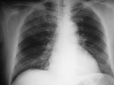
 The new england journal of medicine
pressure (in five of nine) were common. Oxygensaturation while breathing room air was less than
95 percent in seven of nine patients. Physical exam-ination was normal in all patients outside the respi-ratory system. Crackles were noted at the basessymmetrically (in three of nine patients) or asym-metrically (in two of nine). Bronchial breath soundsor egophony were noted in three patients. Chest ra-diographs revealed abnormalities in all of the ninepatients who underwent radiography. In all butthree patients, changes were bilateral and predomi-nantly in the basal lung zones. Abnormalities weresubtle at first in five of the nine patients, primarilyinvolving a reticular interstitial pattern. For two of
these patients, subsequent chest radiographs wereread as normal. All other patients had progressivesymmetric involvement of predominantly air-spacedisease on subsequent radiographs. Pleural effu-sions were not seen. A representative series of chestradiographs is shown in Figure 3.
The new england journal of medicine
pressure (in five of nine) were common. Oxygensaturation while breathing room air was less than
95 percent in seven of nine patients. Physical exam-ination was normal in all patients outside the respi-ratory system. Crackles were noted at the basessymmetrically (in three of nine patients) or asym-metrically (in two of nine). Bronchial breath soundsor egophony were noted in three patients. Chest ra-diographs revealed abnormalities in all of the ninepatients who underwent radiography. In all butthree patients, changes were bilateral and predomi-nantly in the basal lung zones. Abnormalities weresubtle at first in five of the nine patients, primarilyinvolving a reticular interstitial pattern. For two of
these patients, subsequent chest radiographs wereread as normal. All other patients had progressivesymmetric involvement of predominantly air-spacedisease on subsequent radiographs. Pleural effu-sions were not seen. A representative series of chestradiographs is shown in Figure 3.