Pediatr Nephrol (2010) 25:169–172DOI 10.1007/s00467-009-1284-9
Cellular stress-response modulators in the acute rat modelof peritoneal dialysis
Michael Boehm & Helga Bergmeister &Klaus Kratochwill & Regina Vargha &Hans Lederhuber & Christoph Aufricht
Received: 9 February 2009 / Revised: 25 May 2009 / Accepted: 15 June 2009 / Published online: 25 August 2009# IPNA 2009
Abstract Cytotoxicity of peritoneal dialysis fluids (PDF)
Keywords Heat-shock proteins . HSP-72 .
not only results in cellular injury, but also induces heat-
Peritoneal dialysis . Indomethacin . Quercetin
shock proteins (HSP), the main effectors of the cellularstress response. This study investigated effects of modula-tion of mesothelial HSP expression on peritoneal membrane
integrity during acute PDF exposure. In the acute in vivorat model of peritoneal dialysis (PD), either the HSP
Peritoneal dialysis (PD) is a safe, cost-effective, and widely
coinducer indomethacin or the HSP suppressor quercetin
used form of renal replacement therapy in children with
was added to standard PDF (CAPD 3, Fresenius, Ger-
end-stage renal failure. However, PD fluids (PDF) are not
many). HSP-72 expression, number of detached mesothelial
biocompatible, and exposure of mesothelial cells to PDF
cells, and peritoneal protein loss were evaluated at the end
results in marked cytotoxicity []. Recently, we and others
of a 4-h dwell time. Compared with pure PDF exposure,
have demonstrated that cytotoxicity of PDF not only results
addition of indomethacin resulted in increased expression
in cellular injury in experimental models of PD, but also
of mesothelial HSP-72, reduced mesothelial cell exfolia-
induces cytoprotective heat-shock proteins (HSP)
tion, and reduced peritoneal protein loss. Addition of
Induction of HSP-72, the main effector of the mammalian
quercetin resulted in decreased expression of HSP-72,
cellular stress response, was found in mesothelial cells in in
increased mesothelial cell exfoliation, and higher peritoneal
vitro, ex-vivo, and in vivo PDF exposure systems.
protein loss. Differences were statistically significant
Pretreatment by heat or PDF and transfection of HSP-72
between indomethacin-treated and quercetin-treated rats.
results in cytoprotection of mesothelial cells against PDF
Mesothelial HSP expression was related to markers of
exposure, and heat pretreatment of rats prevented mesothe-
peritoneal membrane integrity upon in vivo PDF exposure,
lial cells from detachment from their peritoneal lining in the
consistent with HSP-mediated cytoprotection. These data
in vivo model of PD [, ]. Although these findings suggest
clearly demonstrate the potential for clinically feasible
that HSP-mediated cytoprotection might be an attractive
pharmacologic interventions with the cellular stress re-
novel approach to reduce PDF-mediated peritoneal injury,
sponse as a novel therapeutic approach to improve PD
neither of these pretreatments will ever be feasible in the
clinical setting. In this study, we therefore investigated theeffects of pharmacologic manipulation of the mesothelialHSP expression on peritoneal membrane integrity duringacute in vivo PDF exposure.
M. Boehm : H. Bergmeister : K. Kratochwill : R. Vargha :H. Lederhuber : C. Aufricht (*)Department of Pediatric and Adolescent Medicine,
Medical University of Vienna,AKH Wien, Waehringer Guertel 18-20,
Standard chemicals were purchased from Sigma-Aldrich
1090 Vienna, Austriae-mail: [email protected]
(St. Louis, MO, USA) if not specified otherwise. Animal
protocols were approved by the institutional committee on
using the Mann-Whitney U test. Differences were consid-
animal research. For the acute in vivo model of PD [],
ered to be significant given a p<0.05.
male Sprague Dawley rats (average weight 310 g) under-went anesthesia (ketamine 100 mg/kg, 5 mg/kg xylazine,intramuscularly) and were placed on a heated small-animal
operating table. A sterile catheter was inserted into theperitoneal cavity through a small abdominal midline
As shown in Fig. (upper panel), addition of cellular stress-
incision. In three independent experiments on separate
response modulators to PDF had significant effects on HSP
days, a total of 18 rats (six per treatment group) were
expression of mesothelial cells lining the peritoneal cavity in
slowly infused with 35 ml of conventional, single-chamber
the acute rat model of PD. Compared with pure PDF
bag, acidic pH, L-lactate-buffered PDF with 4.25%
exposure, addition of indomethacin resulted in a 38±26%
(236 mmol/L) D-glucose, and an osmolarity of 511
increase of HSP-72 in isolated mesothelial cells, whereas
mosm/L (CAPD3, Fresenius Medical Care, Germany)
addition of quercetin resulted in a 42±29% reduction.
either without additive or with quercetin (4 mg/kg) or with
As shown in Fig. (middle panel), these effects on HSP
indomethacin (50 μM) in 45–60 s. The animal was gently
expression were associated with effects on mesothelial
moved, a small volume of peritoneal fluid aspirated, the
cellular outcome. Compared with pure PDF exposure, the
catheter withdrawn, and the abdomen sutured. Animals
extent of mesothelial cell exfoliation was reduced from
awoke within 20 min after the procedure and had free
median 92 to 42 cells (by 52±36%) with addition of
access to food and tap water. At 4 h after the intraperitoneal
indomethacin, whereas addition of quercetin resulted in a
injection, animals were again anesthetized, sacrificed by
143±58% increase to 241 cells. Addition of cellular stress
cardial puncture and exsanguination, the abdomen opened
response modulators also had effects on peritoneal protein
by a midline incision, and the complete intraperitoneal fluid
loss as a functional parameter of peritoneal membrane
collected. The volume was recorded and total cell count and
integrity. Protein loss in the peritoneal effluent was also
mesothelial cell counts assessed by hand count after
reduced from median 52 to 47.5 mg by 4±3% with
Giemsa staining and by machine count by a coulter counter.
indomethacin and increased by 10±7% to 55 mg with
To assess peritoneal membrane integrity, amount of
quercetin (Fig. lower panel). Differences in these
ultrafiltration, total number of detached mesothelial cells
parameters became statistically significant when compared
and peritoneal protein loss were then computed for each rat
between indomethacin-treated and quercetin-treated rats.
from dialysate samples obtained at the end of the dwell
There were no differences in peritoneal ultrafiltration
time. Mesothelial cells were isolated by a 20-min peritoneal
between groups (indomethacin: 10.5±2.1 ml; quercetin:
washout with 20 ml phosphate-buffered saline (PBS)
10.7±3.8 ml; PDF=100%, 10.6±3.2 ml).
containing 0.1% trypsin and 0.1% ethylenediaminetetraac-etate (EDTA)
For Western blotting, equal amounts of protein samples
from isolated mesothelial cells (5 μg/lane) were separatedby standard sodium dodecyl sulfate polyacrylamide gel
The results of this study clearly confirm the link between
electrophoresis (SDS-PAGE) using a Multiphor II unit (GE
HSP expression and cellular outcome in mesothelial cells
Healthcare, Uppsala, Sweden). Size-fractionated proteins
during in vivo PDF exposure. Our data show potential for
were transferred to polyvinylidene fluoride (PVDF) mem-
pharmacologic interventions to induce HSP-mediated cyto-
branes by semidry transfer in the Multiphor II Novablot
protection against PDF-induced cellular injury in mesothe-
unit (GE Healthcare). Membranes were blocked with 5%
lial cells. Molecular chaperones, with HSP being the
dry milk in Tris-buffered saline Tween-20 (TBST) buffer.
prototype, constitute a large family of soluble proteins that
Membranes were incubated with the primary anti-HSP-72
are found throughout the mesothelial cell; the amount of all
antibody (SPA 810, Assay Designs/Stressgen, Ann Arbor,
HSP isoforms in some instances can constitute up to 5% of
MI, USA). Detection was accomplished by incubation with
total cellular proteins These proteins are known to
secondary peroxidase-coupled antibodies (Sigma-Aldrich)
cooperate in transport and folding of proteins, without
and enhanced chemiluminescence (Western Lightning
altering their own structure, by binding to hydrophobic,
reagent, Perkin Elmer, Boston, MA, USA). Densitometry
normally hidden domains of immature or denatured
was performed with the image analysis software Quanti-
proteins. They prevent disruption of cytoskeletal structures
tyOne (Bio-Rad, Hercules, CA, USA). Differential expres-
and might thus stabilize the mesothelial cell monolayer
sion was derived from the ratio of specific signals in the
linear range of the protein/signal-intensity relationship.
As detachment of mesothelial cells likely represents the
Values for different treatment conditions were compared
morphologic correlate of impaired peritoneal membrane
Fig. 1 Effects of pharmacologic modulation of heat-shock protein-72
(HSP-72) expression on peritoneal membrane integrity in the acute ratmodel of peritoneal dialysis. Western analysis and densitometry of ratmesothelial cells harvested by trypsin peritoneal washout after a 4-hdwell showed increased HSP-72 expression with the HSP coinducerindomethacin (dark gray) and decreased HSP-72 expression with theHSP suppressor quercetin (light gray) compared with pure peritonealdialysis fluid (PDF) (white) (blot and upper panel). Increased HSP-72expression in mesothelial cells upon indomethacin addition to PDFwas associated with significantly lower detachment (middle panel) anddecreased protein loss into the dialysate effluent (lower panel). Dataare shown as box (giving the 25th and 75th percentile), whiskers(giving the 10th and 90th percentile), and median plots. Data wereobtained in six rats in each group in three independent experiments
mesothelial cells indeed resulted in significant cytoskeletalstabilization of mesothelial cell against PDF exposurefollowing heat pretreatment by whole-body hyperthermiaAs heat pretreatment represents no practical therapeuticoption in the clinical setting of PD, our study introducednonstressful interventions with HSP expression during PDFexposure, using well-accepted modulators of the cellularstress response [].
The nonsteroidal anti-inflammatory drug (NSAID) indo-
methacin is known as coinducer of the stress response andthus to enhance cytoprotection [Albeit the detailedcellular mechanisms are still under investigation, indometh-acin is thought to potentiate trimerization and binding of theheat-shock transcription factor-1 (HSF-1) to DNA, therebyincreasing HSP transcription upon additional cellular stressIn contrast, the bioflavonoid quercetin is known assuppressor of the stress response and thus to inhibit HSP-mediated cytoprotection ]. Quercetin is thought toinhibit hyperphosphorylation and binding of HSF-1 toDNA, thereby decreasing HSP transcription upon addition-al cellular stress In previous studies, indomethacintreatment resulted in attenuation, whereas quercetin treat-ment resulted in aggravation of organ damage in experi-mental disease models, depending on their effects on HSPexpression ].
In our study in experimental PD, pharmacologic modu-
lation of the mesothelial-cell stress response also resulted inconcordant effects on HSP-72 expression and peritonealmembrane integrity, assessed by mesothelial-cell detach-ment and peritoneal protein losses. As expected, addition ofthe coinducer indomethacin to PDF increased, whereasaddition of the suppressor quercetin decreased HSPexpression in mesothelial cells [–Consistent withthe concept of HSP-mediated cytoprotection, the higher
integrity, such HSP-mediated cytoprotection might be an
expression of HSP was clearly reflected in reduced
attractive novel approach to reduce technical failure of PD
mesothelial-cell detachment from its monolayer lining the
. Based on this concept, we recently studied the
peritoneal cavity during the in vivo PDF dwell [–In
relationship between PDF-induced cytoskeletal disruption
addition, we found evidence for an attenuated peritoneal
and HSP-mediated cytoskeletal repair. In the acute rat
barrier dysfunction, as demonstrated by a lower protein
model of PD, overexpression of HSP in peritoneal
content in the PD effluent of indomethacin-treated rats.
Certainly, future studies in a chronic rat model of PD are
fluids induce the stress response in human mesothelial cells. Perit
needed to investigate chronic effects of modulation of
4. Bender TO, Witowski J, Aufricht C, Endemann M, Frei U,
mesothelial HSP expression during chronic PDF exposure.
Passlick-Deetjen J, Jorres A (2008) Biocompatibility of a
With the resulting data, we could then assess the biological
bicarbonate-buffered amino-acid-based solution for peritoneal
relevance of our limited acute findings and extend the data
with regard to peritoneal fibrosis, neoangiogenesis, and
5. Ruffingshofer D, Endemann M, Arbeiter K, Bidmon B, Mueller T,
Herkner K, Aufricht C (2003) Induction of heat shock protein 72
ultrafiltration as important outcome variables. Taken to-
in mesothelial cells exposed to peritoneal dialysate effluent. Perit
gether, our acute experiments extend the previous findings
of HSP-mediated cytoprotection of mesothelial cells fol-
6. Arbeiter K, Bidmon B, Endemann M, Bender TO, Eickelberg O,
lowing heat pretreatment to a more feasible pharmacolog-
Ruffingshofer D, Mueller T, Regele H, Herkner K, Aufricht C(2001) Peritoneal dialysate fluid composition determines heat
ical intervention model. This study suggests potential for
shock protein expression patterns in human mesothelial cells.
cytoprotective additives to PDF to optimize cellular
responses to pathophysiological stress upon PDF exposure.
7. Bidmon B, Endemann M, Arbeiter K, Ruffingshofer D, Regele H,
Data such as ours represent a first step to innovative
Herkner K, Eickelberg O, Aufricht C (2004) Overexpression ofHSP-72 confers cytoprotection in experimental peritoneal dialysis.
therapies to improve the long-term outcome of PD.
8. Endemann M, Bergmeister H, Bidmon B, Boehm M, Csaicsich D,
Acknowledgement This research work was funded by the Else-
Malaga-Dieguez L, Arbeiter K, Regele H, Herkner K, Aufricht C
Kröner-Fresenius Stiftung and by the FWF (Austrian Science Fund)
(2007) Evidence for HSP-mediated cytoskeletal stabilization in
Project P18130-B13 (both to CA). We are grateful to Michaela
mesothelial cells during acute experimental peritoneal dialysis.
Endemann and Klaus Arbeiter for technical assistance and helpful
9. Aufricht C (2005) Heat-shock protein 70: molecular supertool?
10. Gotloib L, Waisbrut V, Shostak A, Kushnier R (1995) Acute and
long-term changes observed in imprints of mouse mesotheliumexposed to glucose-enriched, lactated, buffered dialysis solutions. Nephron 70:466–477
1. Devuyst O, Topley N, Williams JD (2002) Morphological and
11. Westerheide SD, Morimoto RI (2005) Heat shock response
functional changes in the dialysed peritoneal cavity: impact of
modulators as therapeutic tools for diseases of protein conforma-
more biocompatible solutions. Nephrol Dial Transplant 17(Suppl 3):
12. Lee BS, Chen J, Angelidis C, Jurivich DA, Morimoto RI (1995)
2. Arbeiter K, Bidmon B, Endemann M, Ruffingshofer D, Mueller T,
Pharmacological modulation of heat shock factor 1 by antiin-
Regele H, Eickelberg O, Aufricht C (2003) Induction of
flammatory drugs results in protection against stress-induced
mesothelial HSP-72 upon in vivo exposure to peritoneal dialysis
cellular damage. Proc Natl Acad Sci U S A 92:7207–7211
13. Kelly KJ, Baird NR, Greene AL (2001) Induction of stress
3. Aufricht C, Endemann M, Bidmon B, Arbeiter K, Mueller T,
response proteins and experimental renal ischemia/reperfusion.
Regele H, Herkner K, Eickelberg O (2001) Peritoneal dialysis
Tariffe in Euro in vigore dal 18/06/2012. Includono IVA (laddove applicabile), Security Surcharge e Fuel Surcharge*. documenti Per pesi maggiori consultare www.tnt.it oppure contattare il Servizio Clienti TNT(*) A causa delle costanti fluttuazioni del costo del petrolio il Fuel Surcharge potrà subire delle variazioni nei mesi successivi. Per ulteriori informazioni visitate il nostro sito w
IL SINDACO Vista la deliberazione n. 341 del 12.5.1999 con la quale la Giunta comunale, nel precisare che durante i quattro giorni di permanenza nella stalla di Contrada e nelle sei prove di Piazza i cavalli del Palio possono avere la necessità di proseguire eventuali terapie in atto o andare incontro ad alterazioni del normale stato fisiologico che, anche se non di rilevante gravità e tali
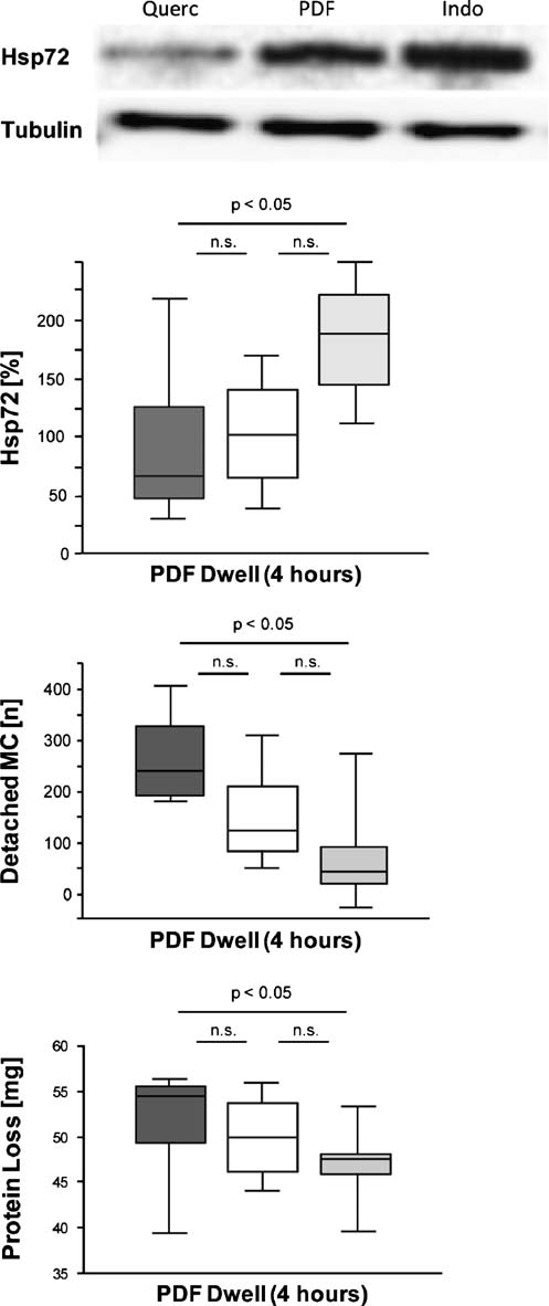 Fig. 1 Effects of pharmacologic modulation of heat-shock protein-72
(HSP-72) expression on peritoneal membrane integrity in the acute ratmodel of peritoneal dialysis. Western analysis and densitometry of ratmesothelial cells harvested by trypsin peritoneal washout after a 4-hdwell showed increased HSP-72 expression with the HSP coinducerindomethacin (dark gray) and decreased HSP-72 expression with theHSP suppressor quercetin (light gray) compared with pure peritonealdialysis fluid (PDF) (white) (blot and upper panel). Increased HSP-72expression in mesothelial cells upon indomethacin addition to PDFwas associated with significantly lower detachment (middle panel) anddecreased protein loss into the dialysate effluent (lower panel). Dataare shown as box (giving the 25th and 75th percentile), whiskers(giving the 10th and 90th percentile), and median plots. Data wereobtained in six rats in each group in three independent experiments
mesothelial cells indeed resulted in significant cytoskeletalstabilization of mesothelial cell against PDF exposurefollowing heat pretreatment by whole-body hyperthermiaAs heat pretreatment represents no practical therapeuticoption in the clinical setting of PD, our study introducednonstressful interventions with HSP expression during PDFexposure, using well-accepted modulators of the cellularstress response [].
Fig. 1 Effects of pharmacologic modulation of heat-shock protein-72
(HSP-72) expression on peritoneal membrane integrity in the acute ratmodel of peritoneal dialysis. Western analysis and densitometry of ratmesothelial cells harvested by trypsin peritoneal washout after a 4-hdwell showed increased HSP-72 expression with the HSP coinducerindomethacin (dark gray) and decreased HSP-72 expression with theHSP suppressor quercetin (light gray) compared with pure peritonealdialysis fluid (PDF) (white) (blot and upper panel). Increased HSP-72expression in mesothelial cells upon indomethacin addition to PDFwas associated with significantly lower detachment (middle panel) anddecreased protein loss into the dialysate effluent (lower panel). Dataare shown as box (giving the 25th and 75th percentile), whiskers(giving the 10th and 90th percentile), and median plots. Data wereobtained in six rats in each group in three independent experiments
mesothelial cells indeed resulted in significant cytoskeletalstabilization of mesothelial cell against PDF exposurefollowing heat pretreatment by whole-body hyperthermiaAs heat pretreatment represents no practical therapeuticoption in the clinical setting of PD, our study introducednonstressful interventions with HSP expression during PDFexposure, using well-accepted modulators of the cellularstress response [].