COMMISSION ON POWDER DIFFRACTION INTERNATIONAL UNION OF CRYSTALLOGRAPHY http://www.iucr.org/iucr-top/comm/cpd/ NEWSLETTER No. 25, July 2001 http://www.iucr.org/iucr-top/comm/cpd/Newsletters/ . IN THIS ISSUE Structure Determination from Powder Diffraction Data (Bill David, Editor) CPD chairman’s message,Paolo Scardi Ab-initio structure determination of oligopeptides from powder diffraction data Editor’s message, Bill David K D M Harris, R L Johnston, E Tedesco and G W TurnerCPD projects: Correlating crystal structure with the physical Quantitative Phase AnalysisRR, Ian Madsen properties of pharmaceutical compounds Size-Strain RR,Davor Balzar N Shankland, W I F David, K Shankland, A Kennedy, CS Frampton and A FlorenceWWW sites related to Powder Diffraction EXPO: New developments IUCr Commission on Powder Diffraction A Altomare, C Giacovazzo, A G G Moliterni and R RizziStructure determination from powder diffraction data News from ICDD and IXAS 26 Revisiting the 1998 SDPD Round Robin Computer Corner,L M D Cranswick What’s On A 117-atom structure from powder diffraction data L B McCusker, Ch Baerlocher and T WesselsCompanies Drug polymorphism and powder diffraction How to receive the CPD Newsletter P Sieger, R Dinnebier, K Shankland and W I F DavidCalls for contributions to CPD newsletter 26 Malaria, synchrotron radiation and Monte Carlo P W Stephens, S Pagola, D S Bohle and A D KosarA case of mistaken identity: metastable Me2SBr2 A N Fitch, G B M Vaughan and A J MoraCombined Rietveld and stereochemical- restraint refinement with high resolution powder diffraction offers a new approach for obtaining protein-drug structures On the reliablility of Rwp in structure prediction Revisiting the 1998 SDPD Round Robin
published now proving that a solution was obtainable frompowder data [5]. Armel Le Bail (1) and Lachlan M.D. Cranswick (2)PARTICIPANTS 1. Université du Maine, Laboratoire des Fluorures,
The 70 people who downloaded data may be considered
CNRS ESA 6010, Avenue O. Messiaen, 72085 Le
to be subscribers to this Round Robin. The possibility was
Mans cedex 9, France - E-mail: [email protected]
given for either anonymous download or filling a Web
2. Lamont-Doherty Earth Observatory of Columbia
form asking for details about which methods and software
University PO Box 1000, 61 Route 9W Palisades, New
will be used for 3 main steps : structure factors extraction,
structure solution and structure completion and refinement. E-mail: [email protected]
31 subscribers filled in the Web form, more or less
INTRODUCTION
completely, indicating that they intended to use some of
In the middle of 1998, the number of structure
the best known programs such as GSAS, FULLPROF,
determinations by powder diffractometry (SDPD) was
SHELX and SIRPOW. 11 participants gave explicit
close to 300 of which 250 were published in the period
answers to all the 3 main steps, simultaneously. One expert
1992-1997 [1]. At that time, a huge number of methods
indicated after the deadline that he would have participated
and computer programs had already proven, at least once,
if the molecular shape had been given for sample 2.
their efficiency in succeeding in the various steps of the
RESULTS AND DISCUSSION
process of solving structures from powder diffraction data.
In the end, we received 5 full questionnaires from 4 final
The word "routine" was pronounced more and more
participants; one questionnaire for sample 1 and four for
frequently, so that it was considered timely to organize a
sample 2. Participant 1 made a very rapid reply but was
Round Robin, in order to try to clarify the various claims
unable to provide coordinates. By a search in the
about the ease or otherwise in performing SDPDs. Data
Cambridge Structural Database, he easily found the
and questionnaires were made available from a Web site
reference for the pharmaceutical compound as being the
starting from May 18, 1998 and the deadline was the last
tetracycline (alias achromycin) hydrochloride. He then
day of June. The competition was spammingly announced
suggested that the coordinates should be found in this
at many Newsgroups and Mailing lists related to
reference. Unfortunately, however, the coordinates were
crystallography and material science. Mails were sent also
not available in this paper or in the Cambridge Structural
to some chemistry lists (Chemweb and CCL), trying to
Database. Only the molecular formula was available.
interest structure predictors to undertake first principles or
Participant 2 was the only regular subscriber to have sent a
semi-empirical calculations. Moreover, personal e-mails
successful questionnaire. He focused his attention
were sent to a number of well-known experts. As a
exclusively on sample 2 and solved its structure, including
consequence of this campaign, more than 800 visitors had
the hydrogen atom positions by the global optimization
a link to the homepage, which is still available [2]. 70 of
method. A model for the molecule was taken from the
the 800 visitors downloaded the data.
tetracycline hydrate in the Cambridge Structural Database
SELECTION OF SAMPLES FOR ANALYSIS
(TETCYH10 entry) and the water was removed. The
There is a clear distinction between compounds for which
tetracycline fragment and the Cl atom were positioned at
prior knowledge is available (molecular formula) or not.
random in the unit cell and an optimum position was
This difference may lead to one choosing quite different
searched (Fig. 1) by simulated annealing using the DRUID
methods for solving the crystal structures. It was thus
program against the 100 first structure factors extracted by
decided to propose two samples that fulfilled these
the Pawley method from the synchrotron data. The final
conditions. We restricted the scope of this Round Robin to
Rietveld refinement plot is shown on the Figure 2. There is
the structure solution part by providing the cell and space
something curious between the starting and final model.
group information. The first sample was inorganic, a
The main move is that O2 and N1 in the TETCYH10
carbonatocobalt(III)pentamine nitrate hydrate; the second
model have rotated by 180° along the C2-C3 axis. The H
sample was organic, the pharmaceutical compoundtetracycline hydrochloride. A medium resolutionsynchrotron pattern was provided for the latter, as well as aconventional X-ray powder pattern with similar resolution. The organic sample was especially selected for modellocation methods; the molecular shape, however, was notgiven. We considered that the shape could have been veryeasily obtained from various sources. During the RoundRobin course, one of the participants gave a very accuratestructure for tetracycline hydrochloride that even includedhydrogen positions. Thus for validation purposes, it wasfound necessary to record a data set from a very smallsingle crystal (40x30x20µ) selected in the powder, usingthe Daresbury 9.8 station equipped with the SMARTSiemens system [3]. The subsequent structure wasdetermined easily (SHELXS) and refined without any
Fig. 1 Comparison of the molecular structures of tetracycline
constraint, including the hydrogen atoms [4]. This raises
hydrochloride obtained from global optimization and
the question of what constitutes a powder and what a
from the final Rietveld refinement (Participant 2)
single crystal sample. The inorganic structure is also
Fig. 3 Final conventional X-ray diffraction Rietveld plot for Fig. 2 Final synchrotron X-ray diffraction Rietveld plot
flood of results? The solving of sample 2 structure from
Patterson is not really the way that most crystallographers
atoms did not moved much between the initial and final
would have expected. Preconceived ideas would have
model. An additional hydrogen atom should have been
prevailed that the unique Cl atom would not have been so
found for building the complete sample 2 structure, O2 in
heavy that a Patterson would have easily disclosed it.
the hydrate becoming an OH. This hydrogen was not
Participant 4 obtained RF=0.57 with the Cl atom.
included by participant 2. Interviewed on this question,
Remember that putting anything at any place gives you
participant 2 commented that the exclusion of the
already RF=0.5 or 0.6. In fact, the structure solution as
hydrogen atom was an oversight caused by no sleep on the
described by participant 4 appears disarmingly simple, but
previous night. The diffraction pattern had been
it is not that straightforward. Here is why. Let us examine
downloaded and the structure solved the day after a trans-
the Fourier difference as Participant 4 provided it. The 2
Atlantic flight. The total time for solution was two hours.
main first peaks are not atoms, neither is the fourth, the
Participant 3 did not have easy Web access and obtained
seventh nor the ninth. Many standard crystallographers
the data by e-mail. He thought that sample 2 would be
would have given up at this stage, but not Participant 4. He
unsolvable without the molecule connectivity and asked
was able to recognize a connected chain of 6 atoms. Here
for it. We had anticipated that we would reply positively to
is the importance of skill, and experience. Most people
such a request, as the connectivity could normally be
would have stopped, rejecting this Fourier synthesis
independently determined by a chemist using other
because of the two first intense peaks do not correspond to
methods such as magnetic resonance. Participant 3 sent
anything, or perhaps would have attempted a refinement of
filled questionnaires for samples 1 and 2, estimating finally
the coordinates, which would have failed. Many would not
that both of them were unsolvable. We are forced to
even have believed that a Fourier synthesis with only the
conclude that the remaining participants found the
Cl atom would have a chance to be successful. The
structures either non-routine, non-solvable or too
organizers did not try the Patterson method because they
had the preconceived idea that it was impossible (in factwe continue to think that way). Because the SDPDRR is
Participant 4 downloaded the data anonymously and
mainly a YES/NO Round Robin (i.e. you win or not), we
solved the sample 2 structure from the conventional X-ray
should take all those lacking questionnaires for 68x2 as a
data by using the CSD package. 158 structure factors were
failure to solve. Perhaps, we should not count the 70 data
extracted by using the CSD-PROFAN program. Using the
downloaded but only the 31 regular subscribers.
CSD-MAIN program, the chlorine atom was located by
Anonymous downloaders never formally declared their
Patterson methods. The first Fourier map produced the
intention to solve the problems. However, it should be
coordinates of ten of the other atoms. Several cycles of
noted that if single crystal data had have been provided,
Rietveld and Fourier syntheses were required to complete
structure solving would have been “routine” using all
the structure (Figs. 3 and 4). According to participant 4, the
freely and commonly available single crystal structure
full time needed for solution and refinement was only 3hours, 2 cups of coffee and 5 cigarettes by using a low-endIntel PC. Participant 4 wrote also that "the structure of theinorganic complex is very simple and that is why it is notinteresting."
It should be stated that participant 2 had provided the
most accurate results with mean displacements relative tothe single crystal data lower by a factor 2 than those fromparticipant 4 and from the organisers [2]. Even thehydrogen atom positions were well located with a meanerror of 0.2 angstroms. COMMENTS
If the structure was in fact quite simple to solve using
Fig. 4 Tetracycline hydrochloride model built from Patterson
Patterson - doesn't it say something t hat there was not a
and Fourier recycling (Participant 4).
solution packages; e.g.; SHELXS, SIR, DIRDIF, CRUNCH.
approaching 500, and the proportion of organic compoundsslightly increases, but remains lower than 20%. New
CONCLUDING REMARKS AND RECOMMENDATIONS
programs for molecule location have been made available
The conclusion from this 1998 Round Robin is that
[8]: POWDERSOLVE (having proposed a post-deadline
solving structures “on demand” from powder diffraction is
contribution [9]), PSSP, ENDEAVOUR, TOPAS, ESPOIR,
non-routine and non-trivial, requiring much skill and
etc, or new options of old programs (the upcoming version
tenacity on the part of practitioners (though this should be
of EXPO2000 and the renamed DASH, which was formally
tempered by the fact that no molecule location program
DRUID. Alas a good number of these programs are
was easily available for free from any website in 1998).
commercial. Moreover, the use of the Internet has grown
Publications stating that structure solution using powder
since 1998 so that if the Round Robin had been proposed
diffraction data is now “routine” (especially from the
in 2001, more participants would have had a chance to
perspective of single crystal practitioners attempting
succeed with both samples. Nevertheless, confirming this
powder diffraction based structure solution) could be
hypothesis needs a new Round Robin to be organized.
considered misleading. Providing inaccurate, rosy reviews
can be counter productive with respect to bringing the field
REFERENCES
into disrepute as being one populated by thecrystallographic equivalent of snake-oil salesmen. The
crystallographic definition of “routine” structure solution is
presently based on the single crystal experience, of one
3. CLRC Daresbury Synchrotron, Station 9.8.
where structures literally solve to near completion at the
click of a button. At present much work can be done to
4. Clegg, W. & Teat, S.J. (2000). Acta Cryst. C56, 1343-
enhance powder diffraction based software to give them
single crystal quality automation and robustness to help
5. Zhu, J.H., Wu, H.X. & Le Bail, A. (1999). Solid State
make structure solution from powder diffraction more an
Science 1, 56-62.
attractive method than it is at present.
6. http://www.cristal.org/iniref/ecm18/ecm18sdpdrr.html7. http://www.chemweb.com/alchem/alchem98/catalyst/cto
A report on the SDPD Round Robin delivered at the ECM-
8. http://www.cristal.org/iniref/progmeth.html#n7
18 congress is still available [6], as well as one written by a
9. http://www.msi.com/materials/cases/an_roundrobin.html
scientific journalist, David Bradley [7]. The number ofdetermined structures using powder diffraction data is now
A 117-Atom Structure from Powder
information about the relative intensities of reflections thatoverlap in 2θ. Diffraction Data Lynne B. McCusker, Christian Baerlocher
Consider the three types of samples (single crystal,
"ideal" powder and textured powder) sketched in two
Laboratory of Crystallography, ETH, Zurich, Switzerland
dimensions in Fig. 1a-c. The textured sample is intuitively
INTRODUCTION
intermediate between a perfectly oriented single crystal,
This is the story of how the structure of the very complex
and a powder with crystallites oriented in all directions,
zeolite UTD-1F, with 117 atoms in the asymmetric unit,
and the corresponding two-dimensional diffraction patterns
could be solved from powder diffraction data[1]. The
support this view. The three reflections highlighted in (a),
structure solution was the culmination of a long period ofmethod development that required not only new dataanalysis software, but also a new way of collecting data [2]. But let us begin at the beginning.
Our research group has a long-standing interest in zeolite
structure analysis, and, because zeolites are rarely availablein the form of single crystals, this has always includeddevelopment of powder diffraction methodology. In oursearch for more powerful approaches to zeolite structuresolution, model calculations reported by Hedel et al.[3]prompted us to consider the possibility of exploitingtexture (preferred orientation of the crystallites). Usually,powder diffractionists go to great lengths to avoid any
Fig 1 Two-dimensional schematic drawings of a specimen
preferred orientation in their samples, because it can
and its diffraction pattern for (a) a single crystal, (b) a
severely distort the intensities in the measured diffraction
powder with randomly oriented crystallites, and (c) a
pattern. However, if the data are collected appropriately,
textured powder. The arrows highlight three reflections
this distortion, which is a function function of the
with similar diffraction angles that are separated in thesingle-crystal pattern, but overlap in the normal powder
orientation of the crystallites in the sample and of the
pattern. The diffraction angle 2θ increases radially
sample in the X-ray beam, can provide additional
from the center of each diffraction pattern.
Drug Information Report NAME: Sample, Martha BIRTHDATE: 1949-04-12 CASE #: s102 LAST REVISED: 2007-09-05 Medication Effects and Interactions The following drugs were reviewed, using one or more online drug information databases (e.g., RxList, Drugs.com, DrugDigest.org., PDR Online, etc.) They were identified as current active medications. zolpidem (Ambien),
Reproduction of Permit Terms and ConditionsFacility ID: 0247040079 Issuance type: Title V Draft Permit This version of facility specific terms and conditions was converted from a database format to an HTML file during an upgrade of the Ohio EPA, Division of Air Pollution Control's permitting software. Every attempt has been made to convert the terms and conditions to look and substantively confo
 COMMISSION ON POWDER DIFFRACTION
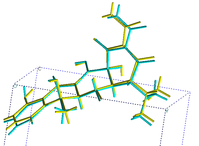 Revisiting the 1998 SDPD Round Robin
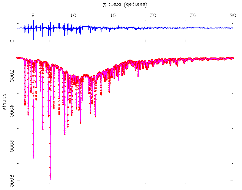
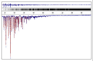
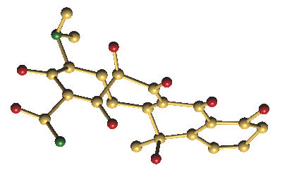 Fig. 3 Final conventional X-ray diffraction Rietveld plot for
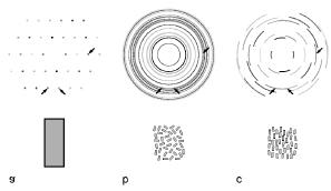 solution packages; e.g.; SHELXS, SIR, DIRDIF, CRUNCH.