Résumé : Reinier de Graaf, physiologiste et histologiste allemand, fut le premier à décire la prostate féminine, à lui attribuer ce terme et à essayer de déterminer la fonction de cet organe. La description qu’en
* Le Federative International Committee on Anatomical Terminology (FICAT) à la conférence d’Orlando (2001) a
fit le gynécologue Américain Alexander J.C.
accepté de mentionner le terme de “prostate féminine” (prostata feminina) dans la nouvelle édition de la
Skene fut l’objet d’un grand débat qui fit
terminologie histologique. Cette décision remplace la terminologie “glandes et canaux para-urétraux de Skene”. ** Des extraits de cette publication ont été présentés au 15th World Congress of Sexology, Paris, 24-28 juin, 2001. passer au second plan les élégantes études d’Huffman. Au début du xxe siècle, la prostate féminine, référencée sous le nom de “glandes
et canaux para-urétraux de Skene – est tou-
et canaux de Skène”, passe pour un vestige
jours utilisé en médecine clinique; dans la
sans importance pour la vie de la femme. La présence de PSA et de paramètres structuraux fonctionnels, ainsi qu’une pathologie, identique à celle de l’homme, Reijnier De Graaf, physiologiste et terminologie anatomique parisienne (Pari- siensia Nomina Anatomica, 1955) on ne men-
tionnait plus les éponymes (Zaviacic, 1999;
le traité De mulierum organis generationi
Zaviacic et Ablin, 2000). Ce n’est qu’en 2001
mettent en évidence qu’il s’agit bien d’un inservientibus… avait pour la première fois
que le FICAT a proposé d’introduire le
équivalent prostatique chez la femme. En 2001 le FICAT* a accepté le terme de prostate et la 4e édition de la terminologie
époque, décrit la structure essentielle de
velle édition de la Terminologie Histologique. histologique la cite comme telle. Les recherches contemporaines montrent
XXe siècle sur la physiologie et la pathologie
que la prostate de la femme a un poids du 1/5
l’urètre féminin. De Graaf avait été le pre-
de la prostate féminine (Evatt, 1911; John-
au 1/4 de celle de l’homme adulte, elle est
mier à essayer de formuler la fonction de
son, 1922; Korenchevsky, 1937; Petrowa et
située dans la paroi de l’urètre et, malgré
cette prostate féminine en écrivant : “La
al. 1939; Caldwell, 1941; Folsom et O’Brien
un espace limité, elle possède un équipement
fonction de la prostate (corpus glandulosum)
1943, 1945; Deter et al. 1946) ont fait pro-
cellulaire muni d’un fonctionnement exocrine
consiste à libérer un liquide glairo-séreux
gressivement évoluer l’opinion sur ce petit
(fluide prostatique féminin), et neuro- endocrine. Par ailleurs, on retrouve les mêmes types et structures cellulaires que
rend plus désirable par son odeur acre et
dans la prostate de l’homme, avec les mêmes
salée, et lubrifie ses parties génitales pen-
fonctions. L’auteur conclut que cela implique
dant le coït” (Jocelyn et al. 1972). Même si
glandes et canaux para-urétraux de Skene)
d’abandonner le terme de glandes de Skène,
sa conception de la prostate féminine était
était considérée comme un organe fémi-
car le terme de prostate féminine est
plus basée sur l’intuition que sur une
nin sans importance, rudimentaire, “ves-
pleinement justifié. La décision du FICAT
recherche effective, De Graaf en est sans
tigial” ne jouant aucun rôle dans la vie de
qui introduit le terme de prostata feminina
doute le découvreur. Par la suite, ce sont
la femme. Sa conception vestigiale était
respecte les nouvelles données de la
basée sur la différence de volume qu’elle
recherche.
biologie, Astruc, Virchow et d’autres, cités
Pour la majorité des médecins, l’origine
(1988) qui se sont intéressés à ce problème
n’était pas un argument suffisant pour
MOTS-CLEFS : KEY WORDS : • Prostate féminine • Female prostate
structures. (Campbell, 1954; Egloff, 1972;
(glandes et canaux (Skene’s para-
domaine reste controversé. Bien que A.J.C. de Skene para- urethral ducts urétraux) and glands)
Skene (1880) ait décrit ces structures 200 ans
Ce qui a permis progressivement d’impo-
• Historique • History
plus tard que De Graaf et que ses conclu-
• Anatomie • Anatomy
sions aient été déjà controversées il y a
• Histologie • Histology
50 ans (Huffman 1948, 1951), jusqu’ici la
d’Huffman (1948, 1951), dont peut consi-
• Ultrastructure • Ultrastructure
prostate féminine est encore connue sous le
dérer qu’ils marquent le début de l’histoire
• Enzymes, PSA • Prostate-specific antigen (PSA)
nom de Skene dans les milieux urologiques
• Enzymes
et gynécologiques; et ce terme – glandes
- VOL.XI, N°41
tate de l’homme; c’est-à-dire des glandes,
1993; Zaviacic et Ablin, 1998, 2000).
Les maquettes en cire de la prostate fémi-
de 150 tubes urétraux de tissus de prostate
Mallon, 1983; Zaviacic et al. 1983; Tepper
nine d’Huffman (1948) restent d’actualité
féminine prélevés lors d’autopsies (Zavia-
et al. 1984). Il y a plus de fibres muscu-
même après 50 ans. Nous avons confirmé
laires lisses dans la prostate féminine que
qu’il existait plusieurs types de prostates
dans celle de l’homme. Les canaux pros-
domaine, dont le début se situe dans la pre-
tatiques (para-urétraux) n’aboutissent pas
l’homme; ce travail a été basé sur l’étude
mière moitié des années 1980, permet de
dans la vulve aux côtés du méat urinaire
d’un matériel d’autopsie beaucoup plus
démontrer que ce petit organe de l’appa-
mais s’ouvrent tout le long de la lumière
reil génito-urinaire de la femme a une struc-
ture, une fonction, et une pathologie défi-
La différence de base entre les prostates
nies. D’autres paramètres comparables à
masculine et féminine réside dans la loca-
avait déjà signalé que le tissu prostatique
lisation du tissu prostatique. Alors que la
féminin était plus riche dans la partie dis-
1999) ont été trouvés progressivement dans
tale de l’urètre, et, plus tard, d’autres
prostatique de l’urètre, chez la femme
auteurs (Zaviacic, 1987, 1999; Wernert et
À présent, les urologues et les gynécologues
c’est tout l’urètre qui lui correspond
al. 1992) l’ont également confirmé. Nous
ont un intérêt grandissant pour cet organe
(Egloff, 1972) (Figure 1). C’est la raison
avons appelé ce type de prostate “type
surtout depuis les nouvelles connaissances
antérieur” (méatal) (Figure 2). Il est pré-
sur l’antigène spécifique prostatique (PSA)
de la prostate féminine. Malgré cela, elle
femmes ; c’est le type le plus fréquent
la prostate masculine, la prostate féminine
(glandes, canaux, musculature lisse) carac-
(Zaviacic, 1987, 1999; Zaviacic et al. 2000).
térisant la prostate masculine, y compris
Eichel et al. (1988) et Eichel (1997) souli-
chez la femme (Zaviacic, Ablin, 1998, 2000).
le “matériel” cellulaire, enzymatique etc.
gnent l’importance du type méatal de la
De même, la pathologie de la prostate fémi-
prostate féminine pour l’obtention de l’or-
nine (carcinome, hyperplasie bénigne pros-
gasme coïtal car, pendant les mouvements
tatique, prostatite, prostatisme) (Zaviacic,
Zaviacic et Whipple, 1993; Zaviacic et al.
coïtaux la partie antérieure de l’urètre
1999), ainsi que d’autres données cliniques
1997). Ces fonctions, surtout l’exocrine,
féminin est stimulée par la pression et les
en rapport avec sa fonction (aspects sexo-
logiques, médico-légaux, gynéco-urolo-
divers champs médicaux (Zaviacic et al.
Plus tard, Eichel (l997) a attiré notre atten-
giques, chronobiologiques) intéressent les
1985; Zaviacic, 1987; Zaviacic et Whipple,
tion sur le point de Gräfenberg (point G),
cliniciens (Zaviacic, 1999). Pendant ces 20 dernières années, unegrande activité de publication a changénotre opinion sur ce petit organe fémi-nin, et on en a gagné progressivementdu terrain en matière de comparaisonavec la prostate masculine (Zaviacic,1999). Nous espérons par nos travaux deces 17 années passées avoir contribué àl’acquisition des connaissances dans cedomaine (Zaviacic, 1999).
Le poids moyen de la prostate d’unefemme adulte est 5,2 g (Zaviacic et al. 2000) et elle représente ainsi à peu près1/5 à 1/4 du poids moyen de la prostatenormale (23,7 g) d’un homme adulte(Thackray, 1978 ; Williams et Warwick,1980; Petersen, 1994). Figure 1 : Le tissu de la prostate féminine dans la paroi de l´urètre féminin (U).
Sur le plan histologique, elle est composée
Une relation entre l’urètre féminin (U), le tissu prostatique, le septum urétro-
des mêmes éléments que ceux de la pros-
vaginal (UVS) et le vagin (V) est démontrée. - VOL.XI, N°41
cologie et en urologie (Kurman, 1994). Al’examen gynécologique et urologique,nous n’avons observé en aucun cas l’exis-tence de canaux séparés de part etd’autres du méat urinaire. Selon notreexpérience, c’est seulement chezquelques multipares, dont l’orifice urétralest élargi, que l’on peut voir – et seule-ment dans la lumière au début de l’urètre– de petits orifices des canaux de la gran-deur de petits points. Selon nos obser-vations, les canaux de la prostate fémi-nine pénètrent dans la lumière urétrale,en arrière du méat de l’urètre et dans toutson trajet. C’est ainsi que l’avaient déjàformulé Huffman (1948) et après luid’autres auteurs, dont nous mêmes,(Zaviacic et al. 1983, 1985; Zaviacic, 1987,1999 ; Wernert et al. 1992). Des travauxcliniques qui corroborent ces dires, sontconsacrés à l’urétroplastie et à l’urétro-lyse externe, au traitement du syndromeurétral et à la correction de la résistance
Figure 2 : Le type “antérieur (méatal)” de la prostate féminin selon la
distale urétrale, causes de dysurie chez la
maquette en cire de Huffman (26).
zone érogène de la paroi antérieure du
(Zaviacic, 1999; Zaviacic et al. 2000). Notre
son et Stonington, 1969). Selon l’opinion
taux d’identification est plus élevé que
chez d’autres auteurs; 66,7 % pour Wer-
“Le type postérieur” de prostate féminine,
nert et al. (1992), 70 % chez Tepper et al.
caractérisé par la présence de tissu pros-
(1984), près de 80 % pour Pollen et Drei-
tatique plus riche dans l’urètre postérieur
linger (1984); Sesterhenn et al. (1998).
vers le col vésical, n’a été retrouvé que
D’autres types – “moyen” ou en forme
dans 10 % de nos études sur pièces d’au-
d’”haltère” – qui ont été décrits, n’ont pro-
contenu à travers l’urètre par un méca-
bablement pas d’importance pratique.
de cas relativement faible qu’il pourrait
Leur fréquence est moins élevée que celle
et localisation de tissu prostatique féminin.
cic et al. 2000). Il faut encore une fois sou-
Le type de prostate “urètre total” n’a été
ligner que le type “antérieur (méatal)” est
par l’urètre que sont excrétées urine et
trouvé que dans 6 % de nos cas d’autop-
la configuration la plus fréquente (66 %
sie (Zaviacic, 1999; Zaviacic et al. 2000).
avec CI 58-74 %). Les types méatal et pos-
On a constaté la présence de cellules sécré-
térieur (10 %) sont présents dans 3/4 de la
toires en microscopie électronique parmi
les cellules des glandes prostatiques fémi-
modèle classique de prostate féminine.
La forme “rudimentaire” de la prostate
était dans notre matériel caractérisée par
canaux de la prostate féminine reste tou-
(Fisher et Jeffrey, 1965; Brandes, 1974; Sri-
l’absence de glandes et de canaux sur la
jours d’actualité. Les données recueillies
gley et al. 1988). Leur cytoplasme apical
plupart des coupes de l’urètre examinées
à partir de l’examen de 250 malades de la
contient des éléments sécrétoires nom-
et elle représente 8 % de nos cas (Zaviacic,
Deuxième clinique gynécologique et obs-
breux (vacuoles et granules sécrétoires),
1999; Zaviacic et al. 2000). Mais, l’examen
tétricale de Bratislava (en Slovaquie), ne
l’urètre sur toute sa longueur, a toujours
Skene (1880), selon qui la prostate fémi-
une configuration sécrétoire active de ces
chant séparément à l’entrée de l’urètre
cellules avec un type de sécrétion apo-
Si on admet qu’une telle constatation équi-
féminin, ce qui est faussement mentionné
crine et mérocrine (Zaviacic et al. 2000).
vaut à identifier une prostate, alors toutes
jusqu’à présent dans la littérature ana-
Entre les cellules sécrétoires et la mem-
les femmes en ont une, quelle que soit sa
forme, même rudimentaire. Si on élimine
basales (des réservoirs). Leur cytoplasme
dans la littérature spécialisée en gyné-
- VOL.XI, N°41
mais sans éléments sécrétoires (Zaviacic et
génicité semblable entre la prostate de
à l’actuelle conception non-vestigiale de la
al 2000). En dehors de deux types de cel-
l’homme et les glandes para-uréthrales de
prostate féminine; organe génito-urinaire
lules prostatiques, on a aussi trouvé des
Skène, comme le montrent la positivité du
cellules de transition placées entre les cel-
Cette période commence à partir de la fin
lules basales et les cellules sécrétoires ou
tatique spécifique). L’expression d’anti-
à proximité. La présence de cellules de
gènes hautement spécifiques à la prostate
d’Huffman (1948, 1951) et continue par des
transition confirme le rôle des cellules
masculine justifie donc l’utilisation du
travaux du début des années quatre-vingt
basales (rôle de réservoir) pour le renou-
jusqu’à nos jours. C’est dans les cinquante
dernières années qu’il y a eu plus particu-
prostate féminine (Zaviacic et al. 2000)
lièrement vingt années de recherche sur la
comme c’est le cas pour la prostate mas-
l’homme, la prostate féminine représente
prostate féminine, dont plusieurs résultats
culine (Xue et al. 1998). L’analyse ultra-
la source essentielle de PSA (Zaviacic et al.
sont originaires de notre institut (Zavia-
structurelle de la prostate féminine nor-
2000). Quand des tissus pathologiques pro-
cic 1984, 2001; Zaviacic and Whipple, 1993,
duisent du PSA, la quantité de PSA sérique
2001; Zaviacic et Ablin, 1998, 2000; Zavia-
et urinaire est la somme de la production
A l’aube du troisième millénaire, la pros-
sentent des cellules sécrétoires et basales
anormale de ces tissus pathologiques, tant
tate féminine se présente comme un organe
chez l’homme que chez la femme (Zaviacic,
génito-urinaire à la structure définie, des
(Zaviacic, 1999; Zaviacic et al. 2000).
paramètres ultrastructurels prostatiques
inclus, avec une fonction exocrine et neuro-
rentes, on a pu prouver qu’il y avait dans
endocrine et une pathologie – donc avec
les cellules sécrétoires des glandes de la
des paramètres comparables avec la pros-
est passée de l’époque de Reijnier de Graaf
(De Graaf, 1672) à la période de la concep-
l’homme (Zaviacic, 1984). L’activité sécré-
tion vestigiale des glandes et des canaux
toire des cellules prostatiques féminines
de Skene para-urétraux dits “non-fonctio-
1- BRANDES D. (1974) Fine structure and
nels et sans importance” du fait du rôle
cytochemistry of male sex accessory organs. In :
prostatique féminin s’appuie sur la preuve
controversé joué par le gynécologue amé-
Brandes D. Male Accessory Sex Organs. Struc-
histochimique E-600 de l’estérase sensi-
ricain Alexandre J.C. Skene (Skene, 1880),
ture and Function in Mammals. New York,
tive, du glucose-6-phosphatase et d’autresenzymes (Zaviacic, 1984). Les différencesdans les activités de naphtyle estérase, glu-cose-6-phosphatase et quelques déshy-drogénases de la prostate féminine, del’âge de la procréation jusqu’après laménopause, pourraient montrer qu’il y aune capacité fonctionnelle différente de laprostate pendant la vie d’une femme etune dépendance fonctionnelle éventuellepar rapport aux hormones sexuellesfemelles (Zaviacic et al.1989). L’antigène spécifique prostatique (PSA) estaujourd’hui le marqueur le plus souventutilisé pour identifier le tissu prostatiqueféminin normal et pathologiquement modi-fié (Pollen et Dreilinger, 1984; Tepper et al. 1984; Wernert et al. 1992; Zaviacic et al. 1994; Zaviacic, 1997; Sloboda et al. 1998;Zaviacic et Ablin, 2000). Le PSA estimmuno-histochimiquement localisé dans
Figure 3 : Agrandissement de la prostate féminine normale. Les cellules
la couche apico-superficielle des cellules
basales (CB) se trouvent sur la membrane basale (MB), dans leur proximité on
sécrétoires prostatiques féminines ainsi que
trouve une partie de la cellule intermédiaire (CI). La lumière de la glande est
dans les cellules uro-épithéliales d’autres
limitée par des cellules hautes sécrétoires cylindriques (CS). Dans leur partie
secteurs du système génito-urinaire fémi-
supranucléaire se trouvent de nombreux éléments de sécrétion (vacuoles
nin (Figure 4) (Zaviacic et Ablin, 2000). sécrétoires et granules sécrétoires). Dans la partie superficielle des cellules sécrétoires se trouvent des protubérances apicales avec vacuoles vides. Les
Entre autres évidences, le concept non ves-
noyaux des cellules sécrétoires, à la différence des cellules basales,
tigial d’une prostate féminine est basé à
contiennent des grands nucléoles et une chromatine dispersée. Uranyl
présent sur la démonstration d’une anti-
acétate, lead citrate, x 10000. - VOL.XI, N°41 prostate gland and its reaction to the male sexualcompounds. J. Physiol. 90; 371-76. 19- KURMAN J. (1994) Blaustein’s Patho-logy of the Female Genital Tract. (4th Ed). New York, Springer Verlag. 20- LADAS A.K., WHIPPLE B., PERRYJ.D. (1982) The G-Spot and Other RecentDiscoveries about Human Sexuality. NewYork, Holt, Rinehart and Winston. 21- MALLON R.B. (1983) Prostatic tissuein females (Letter). J. Sex. Educ. Ther. 9; 6. 22- MOORE K.L. (1980) Clinically Orien-ted Anatomy. Baltimore, London, W. Wil-kins Co. 23- PERRY J.D., WHIPPLE B. (1981) Pel-vic muscle strength of female ejaculators :Evidence in support of a new theory oforgasm. J. Sex. Res. 17 ; 22-39. 24- PETERSEN R.O. (1994) The urinaryFigure 4 : Présence modérée ou élevée de PSA dans la partie apicale superficielle des cellules sécrétoires et dans les membranes des glandes tract and male reproductive system. In :
prostatiques féminines. Technique de biotine-streptavidine-peroxydase. Une femme de 19 ans, x 180.
ed. Philadelphia, J.B. Lippincott. 25- PETROWA E.M., KARAEWA C.S.,
San Francisco, London, Academic Press. of the urethra and vagina of the sexes.
BERKOWSKAJA A.E. (1939) The struc-
2- CALDWELL G.T. (1941) The glands ofture of the female urethra. Arch. Gynec. the posterior female urethra. Texas State
Ultrastructure of human normal and neo-
3- CAMPBELL M. (1954) Urology. Phila-
plastic prostate with comments relative toImmunohistochemical identification of pros-prostatic effects of hormonal stimulation intatic acid phosphatase and prostate specific
4- DE GRAAF R. (1672) De mulierum orga-the rabbit. Amer. J. Clin. Path. 44; 119-34. antigen in female periurethral glands. Uro-
nis generationi inservientibus. Tractatusnovus demonstrans tam homines et animaliaThe female obstructing prostate. JAMA; 121;
27- RAUBER A., KOPSCH F. (1929) Lehr-caetera omnia, quae viviparadicuntur, haudbuch und Atlas der Anatomie des Menschen.minus quam vivipara ab ovo originem ducere.The female urethra. The connecting link bet-
28- RICHARDSON F.H. (1969) Externalween the urologist and the gynecologist.urethroplasty in women : Technique and cli-
SOM A.I. (1946) A clinical and pathologicalnical evaluation. J. Urol. 101 ; 719-23. study of the posterior female urethra. J. Urol.
13- GRÄFENBERG E. (1950) The role of
29- RICHARDSON F.H. (1972) Externalthe urethra in female orgasm. Int. J. Sexol. 3;
urethroplasty for treatment of the urethral syn-
6- EGLOFF B. (1972) Pathologische Ana-drome in the female. Br. J. Urol. : 44; 125. tomie der weiblichen Urethra. In :
14- HUFFMAN J.W. (1948) The detailed
Lubarsch O., Henke F., Rössler R., Uehin-
anatomy of the paraurethral ducts in the
O.G. (1969) Urethrolysis and external ure-
ger E. Handbuch der pathologischen Ana-adult human female. Amer. J. Obstet. Gyne-
throplasty in the female. Surg. Clin. North. tomie. VII/4 Weibliche Geschlechstor-
15- HUFFMAN J.W. (1951) Clinical signi-
31- SCHAEFFER J.P. (1944) Morris Humanficance of the paraurethral ducts and glands.Anatomy. A Complete Systematic Treatise.
7- EICHEL E.W. (1997) Coital orgasmdefined by the CAT research. Abstract
GRAAF R. (1972) On the human repro-
MOSTOFI F.K. (1998) Benign prostate,ductive organs. An annotated translation ofprostatic hyperplasia and prostatic carcinomatractatus de virorum organis generationiin female patients. Mod. Pathol. 11 ; 95 A.
8- EICHEL E.W., EICHEL J.D., KULE S. inservientibus (1668) and the mulierum orga-
33- SKENE A.J.C. (1880) The anatomy and
(1988) The technique of coital alignment andnis generationi inservientibus tractatuspathology of two important glands of theits relation to female orgasmic response andnovus (1672). New treatise concerning thefemale urethra. Amer. J. Obstetr. Diss
simultaneous orgasm. J. Sex Marit. Ther. generative organs of women. J. Reprod. Fer-
9- EVATT E.J. (1911) A contribution to the
17- JOHNSON F.P. (1922) Homologue ofdevelopment of the prostate gland in thethe prostate in the female. J. Urol. 8 : 13-34.
(1998) Metastasizing adenocarcinoma of thehuman female and a study of the homologies
18- KORENCHEVSKY V. (1937) The femalefemale prostate (Skene’s paraurethral glands).- VOL.XI, N°41 Histological and immunohistochemical pros-Update on the female prostate and the phe-tate markers studies and first ultrastructuralnomenon of female ejaculation. J. Sex. Res. observation. Path. Res. Pract. 194 : 129-36.
OKUTANI R., KAWAI T. (1997) Immuno-
49- ZAVIACIC M., ABLIN R.J. (1998) Thehistochemical distribution of rabbit polyclonalfemale prostate (Correspondence). J. Natl. antiurinary protein 1 antibody in the femaleSelected ultrastructural aspects of urothelial(Skene’s gland) and male prostate : new mar-and prostatic tumours. Ultrastruct. Pathol.
50- ZAVIACIC M., ABLIN R.J. (1998) Letterker for neuroendocrine cells ? Acta Histo-
to the Editor. J. Urol. 160 : 1441.
36- STIFTER K.F. (1988) Die dritte
51- ZAVIACIC M., ABLIN R.J. (2000) TheDimension der Lust. Das Geheimnis derfemale prostate and prostate-specific antigen.weiblichen Ejakulation. Frankfurt/Main,
Immunohistochemical localization, implicationsof this prostate marker in women and reasons for
MAN K. (1999) The normal female and theusing the term “prostate” in the human female.male breast epithelium do not express prostate-
GELLER S.A. (1984) Homology between the
Invited Review. Histol. Histopathol. 15 :
specific antigen. Preliminary immunohisto-female paraurethral (Skene’s) glands and thechemical observations of autopsy breast tissues.prostate. Arch. Path. Lab. Med. 108; 423-25.
Gen. Physiol. Biophys. 18; Suppl. 1 : 41-4.
38- THACKRAY A.C. (1978) The male repro-Female ejaculation, female prostate and femaleductive organs. In : Symmers W.St.C. Syste-
sexuality : specific components of female gen-
mic Pathology, 2nd ed. Vol. 4. Edinburgh,
der biology. Sexuologia/Sexology; 1 : 12-18
London, New York, Churchill Livingstone.
HOLOMAN I.K., BREZA J. (2000) Weight,size, macro- anatomy and detailed histology ofthe normal prostate in the adult human female :
OBERUCOVA J. (1983) New information onA minireview. J. Histotechnol. 23 : 61-9.
BERGER K. (1992) “The female prostate” :
the paraurethral (Skene’s) ducts and glands inthe female. Bratisl. Lek Listy. 79 : 533-44 (in :
chemical characteristics and significance.
CIC T., HOLOMAN K. (2000) Immunohis-tochemical study of prostate-specific antigen inGray’s Anatomy. Edinburg, London, Mel-
OBERUCOVA J. (1985) The adult humannormal and pathological human tissues with
bourne, New York, Churchill Livingstone. female urethra. Enzyme-histochemical study.special reference to the male and female pros-tate and breast. J. Histotechnol. 23; 105-11.
SCHALKEN J. (1998) Cell kinetics of pros-
BELOSOVIC M., BREZA J. (2000) Ultra-tate exocrine and neuroendocrine epitheliumstructure of the normal adult human femaleand their differential interrelationship : new
M. (1985) The female prostate or Skene’s parau-prostate gland (Skene’s gland). Anat. Embryol. perspectives. Prostate Suppl. 8 : 62-73. rethral glands and ducts? Reasons for retur-
42- ZAVIACIC M. (1984) Enzyme histoche-ning to the original term of de Graaf. Cs. Gyne-
mistry of the adult human female prostate :Milan Zaviacic* ; Tomas Zaviacic** ; hydrolase and dehydrogenase distribution. Cell. Richard J. Ablin*** ; Jan Breza**** ; Karol Holoman**
43- ZAVIACIC M. (1984) Enzyme histoche-Female urethral expulsions evoked by local digi-
*Institut d’anatomie pathologique. mistry of the adult human female prostate : acidtal stimulation of the G-spot : Differences in thephosphatase distribution. Cell. Molec. Biol. response patterns. J. Sex. Res. 24 : 311-18.
obstétricale de la Faculté de médecine
44- ZAVIACIC M. (1987) The female pros-
CAN J., ZAVIACICOVA A. (1988) Weiblichetate : Nonvestigial organ of the female. A reap-Ejakulation klinisch provoziert. Sexualmedi-
praisal. (Letter to the Editor). J. Sex Marit.
58- ZAVIACIC M., PORUBSKY J., sey, États-Unis.
45- ZAVIACIC M. (1997) Prostate-specific
VIERIK J., HOLOMAN I.K. (1989) Enzy-antigen and history of its discovery. Bratisl. mic equipment of the prostate in women of child-bearing age and women after menopase. Com-
Dérer, Bratislava, République slovaque.
46- ZAVIACIC M. (1999) The Human Femaleparative histochemical study. Cs. Gynekol. 54 :
Pr Milan Zaviacic, Prostate. From Vestigial Skene‘s ParaurethralGlands and Ducts to Woman’s Functional Pros-tate. Bratislava, SAP, 1sted. (le livre + CD).
47- ZAVIACIC M. (2001) The human female
BLAZEKOVA J. (1994) The meaning of pros-prostate and its role in woman’s life : sexologytatic markers in orthology of the female pros-implications. Scand. J. Sexol. 4 : 199-211. tate. Bratisl. Lek. Listy; 95 : 491-97 (in Slovak,
- VOL.XI, N°41 The female prostate: history, functional morphologyand sexology implications*Federative International Committee on Anatomical Terminology (FICAT) at the 2001 meeting atOrlando, Fl, USA has agreed to mention the term female prostate (prostata feminina) in thenew forthcoming edition of Histology Terminology. This decision prohibits further use of theterms of paraurethral glands and ducts, or Skene’s glands for designation of prostate in thehuman female. **Parts of this paper have been presented as an Invited Lecture on 15th World Congress ofSUMMARY : Reinier de Graaf, a Dutch physiologist Sexology, Paris, June 24-28, 2001.and histologist, was the first to describe the female prostate and to assign it this term and was also Milan Zaviacic, MD, PhD, DSc is professor of pathology and forensic medicine at the the first who attempted to formulate the function of this female organ. Description of the American
Comenius University Bratislava, Slovak Republic (Slovakia). The results of Pr Zaviacic’s
gynecologist Alexander J.C. Skene became the
research activities have so far been presented in 440 lectures and up to 550 various scien-
subject of considerable debate increasing lack
tific publications, including full papers (245), editorials, research reports, review articles,
of attention and importance to the female prostate
book chapters and contributions to proceedings of scientific congresses, symposia and
in spite of the elegant studies of the American
conferences (40 of them concerning different aspects of the female prostate). Professor
gynecologist Huffman. At the beginning of 20th century
Milan Zaviacic has established the updated non-vestigial concept of the prostate in the
the female prostate was referred to as Skene’s
female. Based on multidisciplinary research, he has presented the female prostate as a func-
para-urethral ducts and glands, as an insignificant
tional genitourinary organ in the female with a specific structure, function and pathology. rudimentary vestigial organ without any importance
He has shown that the female prostate parameters are similar or even identical with
in the life of women. The expression of the prostate- specific antigen in female Skene’s para-urethral glands
those of the adult male prostate. This recent concept has been based on morphological,
and ducts and structural and functional parameters
histochemical, forensic-medical, sexological, gynecological, urological, chronobiologi-
and diseases similar to that of the male prostate have
cal and pathology research. Professor Zaviacic is member of the Advisory Council Board
provided convincing evidence for the existence of the
of the European Society of Pathology (ESP), member of the National Committee of the Slo-prostate in women. Federative International Committee vak Society of Pathologists, member of the National Committee of the Slovak Sexology Society,on Anatomical Terminology (FICAT) at the 2001 meeting
Vice-President of the Slovak Society of Histochemistry and Cytochemistry, and elected mem-
at Orlando, FL, USA has agreed to mention the term
ber of International Academy of Sex Research (IASR). female prostate (prostata feminina) in the new forthcoming edition of Histology Terminology. This decision prohibits further use of the terms of paraurethral glands and ducts, or Skene’s glands for designation of prostate in the human female.
nature, nevertheless he is undoub-tedly the discoverer of the female
The contemporary research presents the female prostate as an organ with inferior parameters (weight, size, functional productivity) if compared with the male prostate, similarly to many other organs in man. Its average weight is 5.2 g, representing 1/5 to 1/4 of the weight of an adult male prostate. The female prostate is situated in the wall of the urethra that limits its size and weight. Despite the smaller space, its cellular
term (De Graaf, 1672). In his work Deequipment furnishes exocrine function (production of female prostatic fluid) and neuroendocrine function. mulierum organis generationi inser-Equally to the male prostate, glands, ducts and smooth vientibus… Reinier De Graaf (1641-
muscle cells (muscle-fibrous tissue) form the female prostate. The structure, including the ultrastructure of secretory (luminal), basal (reserve) and intermediary cells of the female prostate glands, corresponds to the structure of the same cells in glands of the prostate of an adult male. Also the function of these cells is the same as in the male prostate. Both, the basal cells and from them derived intermediary cells, take part in
and ducts around the female urethra. the renewal of exocrine (secretory) cells of the female prostate glands. (Immuno) histochemistry proved activity of lysosomal and prostate- specific acid phosphatase (PSAP) and disclosed the expression of prostate-specific antigen (PSA) in the luminal-apical
the prostate (corpus glandulosum) is
part of the secretory cells of the female prostate. The expression of the highly specific antigen of the male prostate in this female structure implies the necessity to use the unambiguous term “prostate” also in women. This excludes the Skene’s eponym (Skene’s glands) or the histological descriptive term “paraurethral glands and ducts”, still nowadays
fashion during coitus (Jocelyn et al. incorrectly used by some to identify the female prostate. The terminology decision of FICAT with introducting the term “prostata feminina” respects the novel data achieved in the research of the female prostate. - VOL.XI, N°41
prostate concrements “corpora amy-
tigial position of the female prostate. lacea”, which had before been known
ders. The difference in size, in disfavor
also later on less intensive than that of
gen and its potential implications in the
equally significant role (Zaviacic et al. - VOL.XI, N°41
(Zaviacic and Ablin, 1998), in Letter
inflammation of the female prostate. to the Editor of the Journal of UrologyInvited Review in the Journal of His-tology and Histopathology (Zaviacic
cm (height) (Zaviacic et al. 2000). If we
Macroanatomy, functional morphology and sexology implications
(Figure 2), then the weight of the female
of the female prostate
ert et al. 1992, Sesterhenn et al. 1998).
the male and female prostate glands.
lished over the last 20 years, as well as
jects to the area of the posterior urethra
the size of the prostate, which for these
and the neck of the urinary bladder.
of prostatic tissue was localized in this
Journal of the National Cancer Institute
prostate, it possesses all the structural
prostate, Zaviacic, 1987 ; Zaviacic etal. 2000) and this type correspond tothe place of G-spot. Eichel (1997),Eichel et al. (1988) pointed out theimportance of the meatal type of thefemale prostate for achieving coitalorgasm in the female when the ante-rior portion of the female urethra withthe greatest amount of prostatic tis-sue is directly stimulated by pressureand counterpressure of the genitalregions of the male and female. Eichel,thus turned our attention to the vagi-nal introitus where the urethral mea-tus and onset of the anterior urethraare projected. The female prostate possesses histo-logically the same structures as theprostate of the male, i.e. glands, ducts,and smooth musculature. The ductsare more numerous than the glandsand they exceed in number also theducts in the male prostate. The smoothmusculature (musculofibrous tissue)are also more abundant in the femalecompared to the male prostate(Zaviacic, 1987, 1999 ; Zaviacic et al. 2000). The prostatic (paraurethral)
Figure 1 : The female prostate in the wall of the female urethra (U). The relationship is shown between the female urethra with the prostatic tissue, the urethro-vaginal septum (UVS) and the vaginal canal (V). - VOL.XI, N°41
urethra, they rather penetrate into thelumen of the urethra along its wholelength (Huffman, 1948; Zaviacic, 1999;Zaviacic et al. 2000), and it is throughthe urethra and not through separateopenings that the female prostate dis-charges its contents (Zaviacic, 1999 ;Zaviacic et al. 2000). Similarly as the prostate in the male,the female prostate has at least twomain functions : exocrine – productionof female prostatic fluid – and neu-roendocrine function (Zaviacic, 1999). The exocrine function of the femaleprostate is reflected by its particularstructure, including the presence ofsecretory and basal cells with theircharacteristic ultrastructural appear-ance. Tall cylindrical secretory (lumi-nal) cells are the predominant typeboth in the female and male prostaticglands. Apical cytoplasm containsabundant secretory elements (secre-
Figure 2 : “Anterior (meatal)” type of the female prostate according to Huffman’s wax model (1948).
endoplasmic reticulum, developedGolgi complexes and numerous mito-
tially indicative of a varying functional
of woman’s life (Zaviacic et al. 1989).
logically altered prostatic tissue in the
tory and basal cells (Figure 3) (Zaviacic,
tory elements. Their nuclei, unlike those
uroepithelial cells at other sites of the
female prostate, particularly carcinoma.
that they resemble basal (reserve) cells.
do appear (type 2 intermediary cells).
(reserve) cells in the renewal of cells in
- VOL.XI, N°41
female, however, immunohistochem-ically only the production of serotoninby female prostatic neuroendocrinecells has so far been established. Todate we are but at the beginning in thestudy of the female prostate as a fur-ther neuroendocrine organ, a con-stituent part of the diffuse neuroen-docrine system of the woman(Zaviacic, 1999 and references therein;Zaviacic et al. 1997). Our insight into the exocrine functionof the female prostate in producingfemale prostatic fluid is much moredeveloped than our knowledge on itsneuroendocrine function. It is true thatpure prostatic fluid has not yet beenisolated and it has been studied onlyas a component of the female ejacu-late, whose substantial component it
Figure 3 : Magnified overview of a normal gland of the female prostate. Basal cells (BC) rest on the basement membrane (MB). In the lower part a portion of an intermediary cell (CI). The lumen of the gland is lined with tall cylindral secretory cells (CS) containing secretory vacuoles and secretory granules. Protuberances of the apical part of CS contain empty vacuoles. Uranyl acetate and lead citrate, x 10 000.
(Zaviacic, 1999 and references therein).
(Zaviacic, 1999, 2001). On biochemi-cal analysis, the fluid of urethral expul-sions (female ejaculate) was found tohave a significantly higher concentra-tion of components arising from thefemale prostate, namely prostatic acidphosphatase and especially prostate-specific antigen, and a significantlylower concentration of urea and crea-tinine than urine specimens taken fromthe same women (Zaviacic, 1999). Forensic aspects of the female ejacu-late, containing female prostatic fluid,and the female ejaculation phe-nomenon as such concern twoissues : critique of the significance ofthe acid phosphatase test in evidenceof rape in women and the possibility tostudy modes of secretory mechanismsof the female prostate. Details see inbook (Zaviacic, 1999) and in our recent
Figure 4 : Expression of prostate-specific antigen (PSA) in the apical part of cytoplasm of the secretory cells and in membranes of female prostatic glands. 19 years-old female, biotin-streptavidin-peroxidase technique, x 180. - VOL.XI, N°41
vestigial notion of this female organ.
hyperplasia, prostatitis (female urethral
2001). Similarly as in the male, initiation
diseases is still lacking (Zaviacic, 1999
Milan Zaviacic* ; Tomas Zaviacic** ; RichardJ. Ablin*** ; Jan Breza**** ; Karol Holoman**
lation of the clitoris or suprapubic mas-
similar serious “prostatic” diseases,
(Zaviacic, 1999). Expression of the anti-
trigger urethral expulsions (female ejac-
Pr Milan Zaviacic,
tially play a role also in the motivation
of life threatening erotisizing paraphilic
- VOL.XI, N°41
Inventory Listing for "Zest Restaurant Staten Island". Click on the "Buy Now" link to purchase an item. • The Supermarket Epicure: The Cookbook For Gourmet Food At Supermarket Prices Jo ($3.89) - • Junk Food Buffalo Bills Super Bowl XXV Mens M Medium Blue Vintage T-Shirt NEW ($9.99• BRAZILIAN FOOD [9780857850423] - JANE FAJANS (PAPERBACK) NEW ($39.39) - • BRAND NEW! C
Astma Patiënten Vereniging VbbA / LCP Secretariaat: P.Sijpersma, Loëngasterlaan 29, 8604 ZC Sneek. EVIDENCE-BASED = wetenschappelijk bewijs. Verzameling van uitspraken nationaal en internationaal “Slechts 10 % van de chirurgie is evidence-based. Dat is weinig, maar op basis van je ervaring weet je toch dat een bepaalde behandeling de beste is”. Prof. Wagener, oncoloog, tijdens ee
 Résumé : Reinier de Graaf, physiologiste
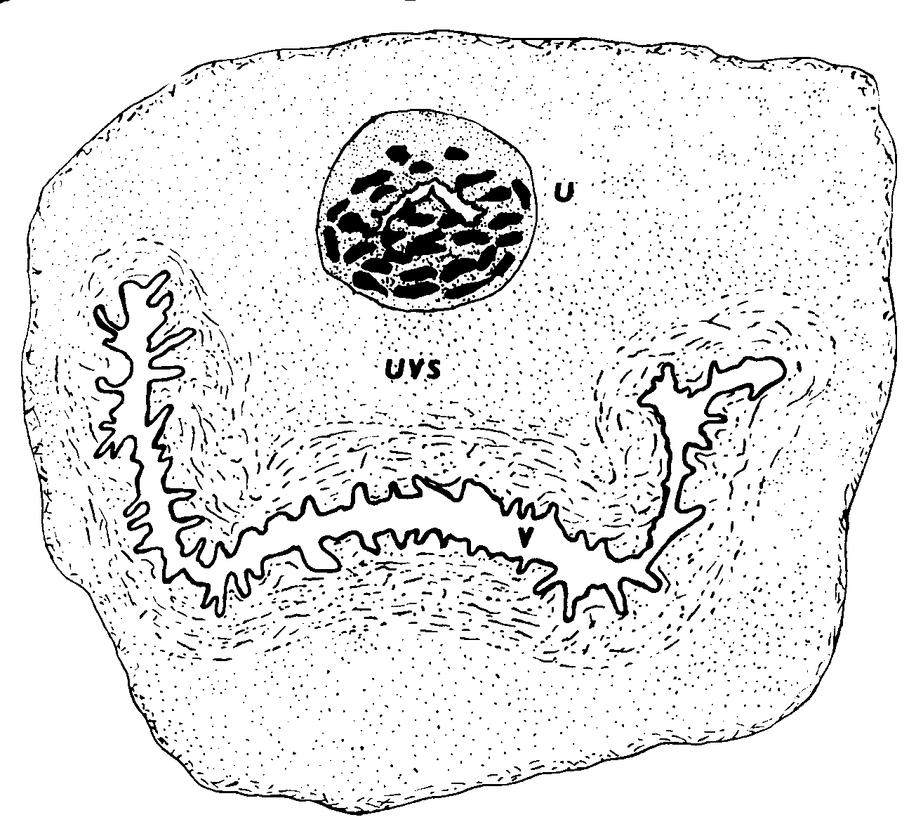 tate de l’homme; c’est-à-dire des glandes,
1993; Zaviacic et Ablin, 1998, 2000).
tate de l’homme; c’est-à-dire des glandes,
1993; Zaviacic et Ablin, 1998, 2000). cologie et en urologie (Kurman, 1994). Al’examen gynécologique et urologique,nous n’avons observé en aucun cas l’exis-tence de canaux séparés de part etd’autres du méat urinaire. Selon notreexpérience, c’est seulement chezquelques multipares, dont l’orifice urétralest élargi, que l’on peut voir – et seule-ment dans la lumière au début de l’urètre– de petits orifices des canaux de la gran-deur de petits points. Selon nos obser-vations, les canaux de la prostate fémi-nine pénètrent dans la lumière urétrale,en arrière du méat de l’urètre et dans toutson trajet. C’est ainsi que l’avaient déjàformulé Huffman (1948) et après luid’autres auteurs, dont nous mêmes,(Zaviacic et al. 1983, 1985; Zaviacic, 1987,1999 ; Wernert et al. 1992). Des travauxcliniques qui corroborent ces dires, sontconsacrés à l’urétroplastie et à l’urétro-lyse externe, au traitement du syndromeurétral et à la correction de la résistance
Figure 2 : Le type “antérieur (méatal)” de la prostate féminin selon la
cologie et en urologie (Kurman, 1994). Al’examen gynécologique et urologique,nous n’avons observé en aucun cas l’exis-tence de canaux séparés de part etd’autres du méat urinaire. Selon notreexpérience, c’est seulement chezquelques multipares, dont l’orifice urétralest élargi, que l’on peut voir – et seule-ment dans la lumière au début de l’urètre– de petits orifices des canaux de la gran-deur de petits points. Selon nos obser-vations, les canaux de la prostate fémi-nine pénètrent dans la lumière urétrale,en arrière du méat de l’urètre et dans toutson trajet. C’est ainsi que l’avaient déjàformulé Huffman (1948) et après luid’autres auteurs, dont nous mêmes,(Zaviacic et al. 1983, 1985; Zaviacic, 1987,1999 ; Wernert et al. 1992). Des travauxcliniques qui corroborent ces dires, sontconsacrés à l’urétroplastie et à l’urétro-lyse externe, au traitement du syndromeurétral et à la correction de la résistance
Figure 2 : Le type “antérieur (méatal)” de la prostate féminin selon la mais sans éléments sécrétoires (Zaviacic et
génicité semblable entre la prostate de
à l’actuelle conception non-vestigiale de la
al 2000). En dehors de deux types de cel-
l’homme et les glandes para-uréthrales de
prostate féminine; organe génito-urinaire
lules prostatiques, on a aussi trouvé des
Skène, comme le montrent la positivité du
cellules de transition placées entre les cel-
Cette période commence à partir de la fin
lules basales et les cellules sécrétoires ou
tatique spécifique). L’expression d’anti-
à proximité. La présence de cellules de
gènes hautement spécifiques à la prostate
d’Huffman (1948, 1951) et continue par des
transition confirme le rôle des cellules
masculine justifie donc l’utilisation du
travaux du début des années quatre-vingt
basales (rôle de réservoir) pour le renou-
jusqu’à nos jours. C’est dans les cinquante
dernières années qu’il y a eu plus particu-
prostate féminine (Zaviacic et al. 2000)
lièrement vingt années de recherche sur la
comme c’est le cas pour la prostate mas-
l’homme, la prostate féminine représente
prostate féminine, dont plusieurs résultats
culine (Xue et al. 1998). L’analyse ultra-
la source essentielle de PSA (Zaviacic et al.
mais sans éléments sécrétoires (Zaviacic et
génicité semblable entre la prostate de
à l’actuelle conception non-vestigiale de la
al 2000). En dehors de deux types de cel-
l’homme et les glandes para-uréthrales de
prostate féminine; organe génito-urinaire
lules prostatiques, on a aussi trouvé des
Skène, comme le montrent la positivité du
cellules de transition placées entre les cel-
Cette période commence à partir de la fin
lules basales et les cellules sécrétoires ou
tatique spécifique). L’expression d’anti-
à proximité. La présence de cellules de
gènes hautement spécifiques à la prostate
d’Huffman (1948, 1951) et continue par des
transition confirme le rôle des cellules
masculine justifie donc l’utilisation du
travaux du début des années quatre-vingt
basales (rôle de réservoir) pour le renou-
jusqu’à nos jours. C’est dans les cinquante
dernières années qu’il y a eu plus particu-
prostate féminine (Zaviacic et al. 2000)
lièrement vingt années de recherche sur la
comme c’est le cas pour la prostate mas-
l’homme, la prostate féminine représente
prostate féminine, dont plusieurs résultats
culine (Xue et al. 1998). L’analyse ultra-
la source essentielle de PSA (Zaviacic et al.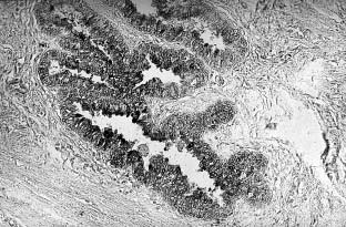 prostate gland and its reaction to the male sexualcompounds. J. Physiol. 90; 371-76.
 The female prostate: history, functional morphologyand sexology implications
*Federative International Committee on Anatomical Terminology (FICAT) at the 2001 meeting at
Orlando, Fl, USA has agreed to mention the term female prostate (prostata feminina) in the
new forthcoming edition of Histology Terminology. This decision prohibits further use of the
terms of paraurethral glands and ducts, or Skene’s glands for designation of prostate in thehuman female.
The female prostate: history, functional morphologyand sexology implications
*Federative International Committee on Anatomical Terminology (FICAT) at the 2001 meeting at
Orlando, Fl, USA has agreed to mention the term female prostate (prostata feminina) in the
new forthcoming edition of Histology Terminology. This decision prohibits further use of the
terms of paraurethral glands and ducts, or Skene’s glands for designation of prostate in thehuman female. (Zaviacic and Ablin, 1998), in Letter
inflammation of the female prostate.
(Zaviacic and Ablin, 1998), in Letter
inflammation of the female prostate.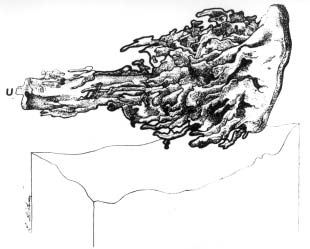 urethra, they rather penetrate into thelumen of the urethra along its wholelength (Huffman, 1948; Zaviacic, 1999;Zaviacic et al. 2000), and it is throughthe urethra and not through separateopenings that the female prostate dis-charges its contents (Zaviacic, 1999 ;Zaviacic et al. 2000).
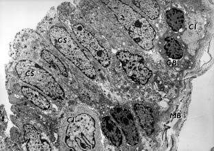
 female, however, immunohistochem-ically only the production of serotoninby female prostatic neuroendocrinecells has so far been established. Todate we are but at the beginning in thestudy of the female prostate as a fur-ther neuroendocrine organ, a con-stituent part of the diffuse neuroen-docrine system of the woman(Zaviacic, 1999 and references therein;Zaviacic et al. 1997).